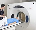
High-field strength magnetic resonance imaging (MRI) help reveal tiny white matter injuries in the developing brain previously undetected using standard MRI.
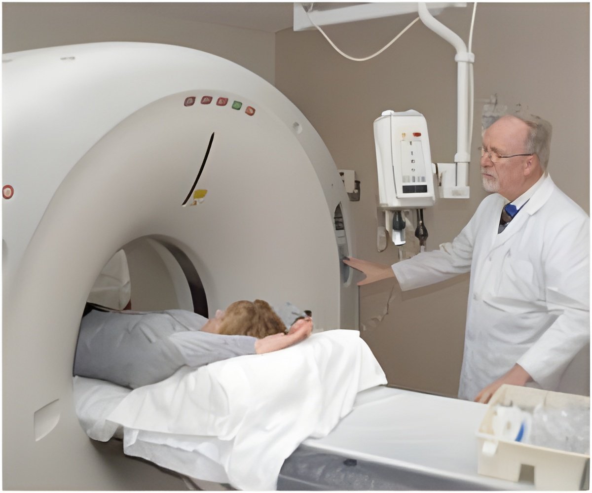
White matter injury is the most common cause of chronic neurologic disability in children with cerebral palsy, explains principal investigator Stephen Back, M.D., Ph.D., but babies with cerebral palsy often have MRIs that miss injury, which creates significant challenges, including delayed treatment intervention and rehabilitation.
"Until now there hasn't been a compelling reason to put preterm babies into a high-field MRI scanner. Our work indicates the magnetic field strength of current clinical MRI may be a limiting factor to detecting some white matter lesions in the preterm infant. Now that we can detect this injury, we also hope our findings may encourage MRI researchers to find more sensitive means to detect this injury with lower field MRIs that are widely available," said Back, an associate professor of pediatrics and neurology in the Papé Family Pediatric Research Institute at OHSU Doernbecher Children's Hospital.
High-field MRI scanners are still mostly used as a research tool and not widely available outside of specialized MRI research centers like OHSU, Back added.
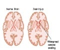
White matter injury occurs during brain development when nerve fibers are actively being wrapped in myelin, the insulation that allows nerve fibers to rapidly transmit signals in the brain. The cells required to make myelin can be easily destroyed when blood flow to the developing brain falls below normal or when maternal infection occurs during pregnancy. The loss of these cells disrupts brain maturation and results in failure to make the myelin required for normal brain function.
Preterm infants are particularly susceptible to these injuries, which can result in lifelong impairments, including inability to walk as well as intellectual challenges.
Advertisements
"Our findings support the potential of using high-field MRI for early identification, improved diagnosis and prognosis of white matter injury in the preterm infant, and our large preclinical animal model provides unique experimental access to questions directed at the cause of these lesions, as well as the optimal field strength and modality to resolve evolving lesions using MRI."
Advertisements
Source-Eurekalert

