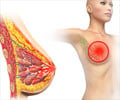
An international team of researchers have developed a new X-ray technique that would allow 3D visualizations of the breast and detect tumours at early stages.
A new X-ray technique could possibly revolutionize the detection of breast cancer, say an international team of researchers. The scientists responsible for this invention claim that this would allow 3D visualizations of the breast and help in early detection of tumors.
Researchers revealed that the new technique known as Analyser-Based X-ray Imaging (ABI), with high spatial resolution, would help in detecting breast tumors with greater precision.Currently, X-ray mammography is the most widely used tool in diagnostic radiology, but it fails to identify about 10 to 20 pct of palpable breast cancers, as some breasts, especially in young women, are very dense. Therefore, on mammograms, glandular tissues can mask cancer lesions.
X-ray computed tomography (CT) could produce accurate 3D images of the entire breast, improving the detection of early diseases in dense breasts. However, its use in breast imaging is limited by the radiation dose delivered to a radiosensitive organ such as the breast.
The new ABI-CT technique has allowed scientists to overcome this problem. They have managed to visualize breast cancer with an unprecedented contrast resolution and with clinically compatible doses.
During the study, the researchers used the new procedure on an in vitro specimen using a radiation dose similar to that of a mammography examination.
The dose corresponded to a quarter of that required for imaging the same sample with conventional CT scanner.
Advertisement
The research team chose a particularly challenging specimen: a breast invaded by a lobular carcinoma (a diffusely growing cancer), the second most common form of breast cancer, which is also very difficult to visualize in clinical mammography.
However, the study showed that high-spatial-resolution ABI-CT makes visible small-size and low-contrast anatomic details that could otherwise only be seen by the microscopic study of an extracted sample of the breast tissue (histopathology).
"We can clearly distinguish more microcalcifications -small deposits of minerals which can indicate the presence of a cancer- than with radiography methods and improve the definition of their shapes and margins", said Jani Keyrilainen, main author of the paper.
"If we compare the images with X-ray mammograms and conventional CT images, we can confirm that this technique performs extremely well", he adds," Jani Keyrilainen added.
The results of this research appear today in the journal ¨Radiology¨.
Source-ANI
TAN/L


