New diagnostic imaging technique could better differentiate benign lung lesions from cancerous ones.
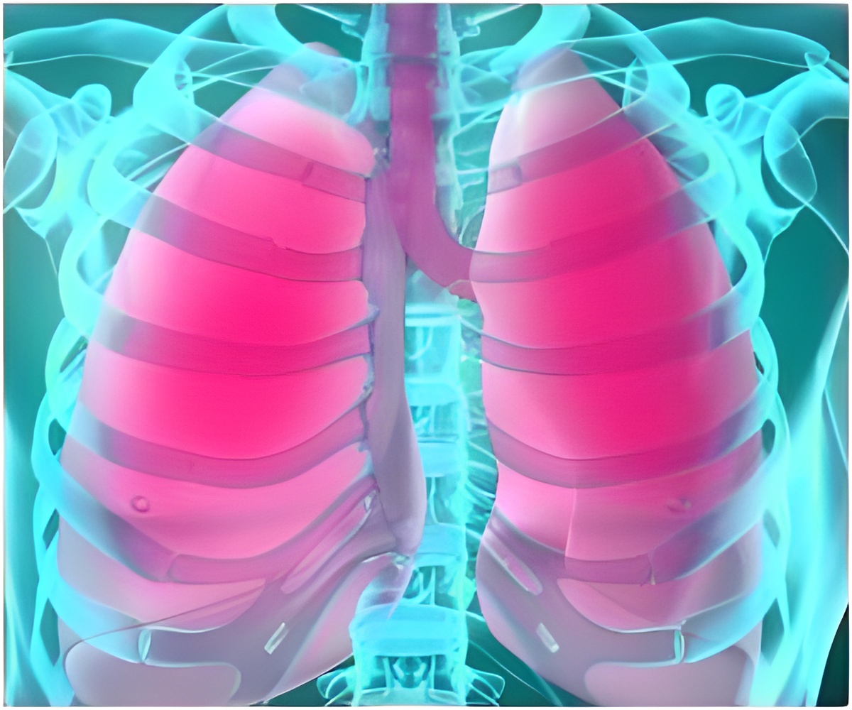
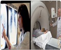
PET-CT scans are currently used by a doctor to determine what stage the cancer is at and whether the detected lung lesions are cancerous. This test involves a CT scan taking pictures from around your body and a PET scan which uses a small amount of an injected radioactive drug to show uptake within structures in your body.
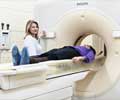
Whilst this is the current gold-standard for treatment, this new research has shown that a type of MRI scan, known as diffusion-weighted MRI, is more accurate. This technique measures water movement in the tissue of the lungs and can detect the structural changes that lung cancer causes, even in the early stages of the disease.
The new technique also has the advantage of being non-invasive and does not require any radiation exposure.
The research analysed 50 people who were due to be operated on and had been diagnosed with lung cancer or suspected lung cancer assessed by PET-CT scan. One day before their operation, the same group also underwent a diffusion-weighted MRI scan.
The results showed that with PET-CT scans, 33 patients were diagnosed correctly, 7 incorrectly and 10 were undetermined. With diffusion-weighted MRI scans, 45 patients were diagnosed correctly and 5 incorrectly. The 10 undetermined cases with PET-CT were correctly diagnosed using diffusion-weighted MRI scan.
Advertisements
"PET/CT scans can wrongly diagnose cancer as they can misinterpret inflammation in the lungs as a malignant lesion. Especially in these inflammatory lesions, diffusion weighted MR is more accurate which could help avoid unnecessary surgical procedures for those people without malignant disease. In addition, it could help to classify patients with lung cancer to enable doctors to provide the most effective therapeutic procedures."
Advertisements
Source-Eurekalert


