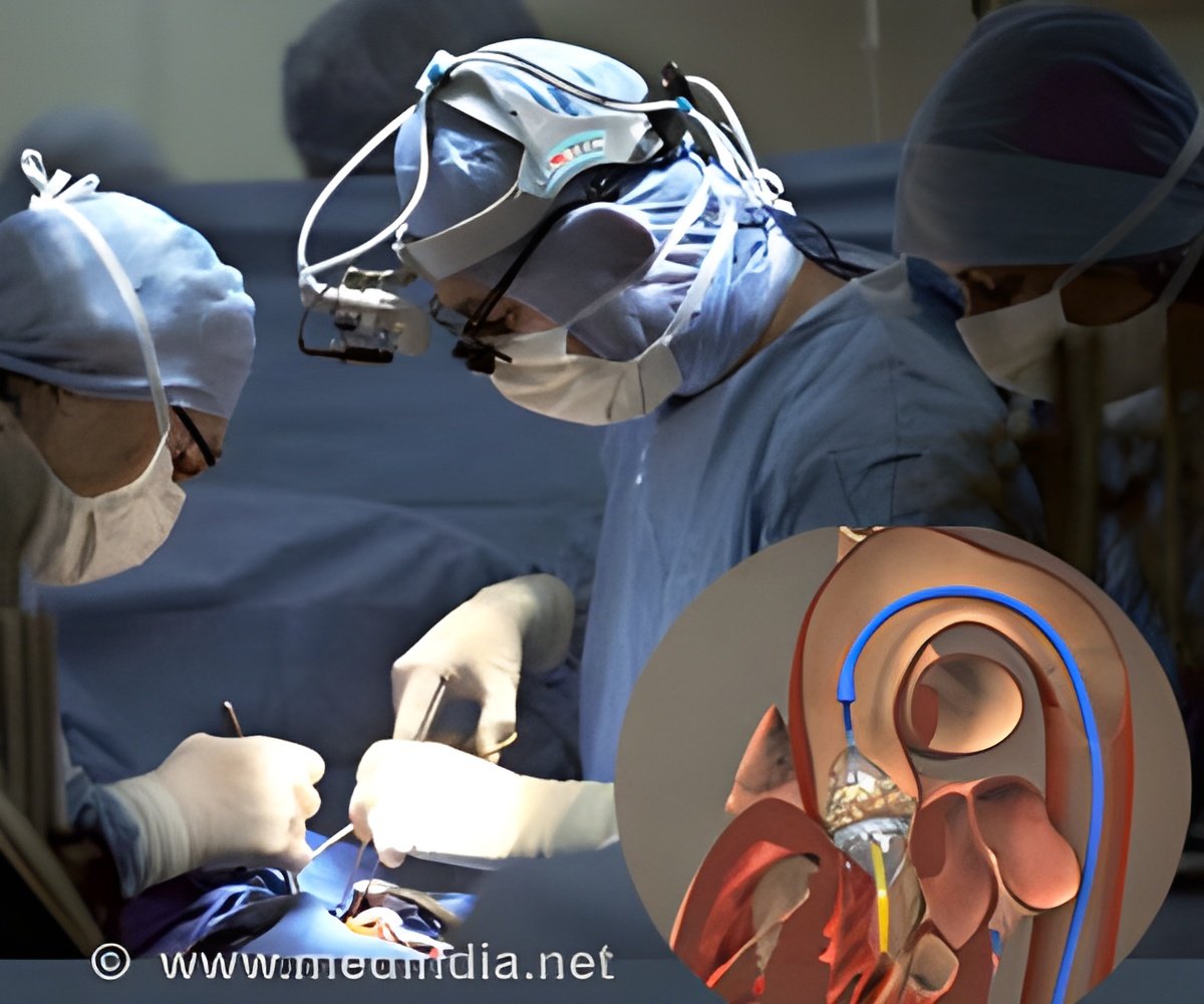
It is possible to observe visually identifiable neuronal networks over a period of several weeks in order to study the after-effects of anesthesia.

‘It is possible to observe visually identifiable neuronal networks over a period of several weeks in order to study the after-effects of anesthesia.’


The awake cortex showed complex activity patterns, with individual cells firing at different times. Under anesthesia, all neurons displayed identical activity patterns and fired at the same time. "While one might expect the brain to cease its activity under anesthesia, in reality, the situation is quite different. Neurons remain highly active but change their communication mode. During unconsciousness they become highly synchonized -- in simple terms all neurons start doing the same thing. " explains Mr. Thomas Lissek, a neurobiologist from Heidelberg and the study's first author. Another surprising finding was that neurons were more sensitive to environmental stimuli under anesthesia than when the brain was awake. "This is especially surprising, as anesthesia is used to block both pain and environmental stimuli during surgery," says Mr. Lissek. Some of the brain regions that are normally dedicated to tactile perception even responded to sound information.
These new insights into neuronal activity patterns provide information regarding the identity of the cellular parameters involved in producing consciousness and the loss of consciousness. Once combined with further advances that would allow us to measure neuronal activity inside the human brain, these findings could contribute to improving our diagnostic capabilities in conditions such as coma and locked-in syndrome.
For the first time, this study succeeded in showing that it is possible to observe visually identifiable neuronal networks over a period of several weeks in order to study the after-effects of anesthesia. "It is clear that investigation of anesthesia will produce deep insights into the mechanism of consciousness", emphasizes Hasan.
Source-Eurekalert


