- Amniotic Band Syndrome - (https://rarediseases.org/rare-diseases/amniotic-band-syndrome/)
- Prenatal diagnosis of amniotic band syndrome - (https://www.ncbi.nlm.nih.gov/pmc/articles/pmc4813076/)
- What is amniotic band syndrome (ABS)? - (http://www.chw.org/medical-care/fetal-concerns-center/conditions/infant-complications/amniotic-band-syndrome/)
- Epidemiology and risk factors of amniotic band syndrome, or ADAM sequence - (https://www.ncbi.nlm.nih.gov/pmc/articles/pmc3530965/)
What is Amniotic Band Syndrome?
Amniotic band syndrome (ABS) refers to various birth defects caused by fibrous bands from the amniotic sac wrapping around various parts of the fetus and preventing normal development.
Other names for the condition include congenital constriction band syndrome (CCBS), Adam complex, and Streeter dysplasia. The condition has been described since the time of Hippocrates and Aristotle (early 300 BC).
Studies have shown the prevalence to be about 7.7/10,000 live births, and as high as 178/10,000 among aborted fetuses.
Fetal Development within the Uterus - In Brief
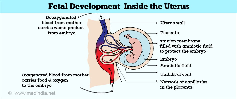
To better understand how amniotic band syndrome occurs, it is necessary to know how a fetus develops in a mother’s uterus. The fetus floats in the mother’s womb surrounded by amniotic fluid. The fluid around the fetus is enclosed by a sac composed of two membranous layers. The outermost membrane is the chorion and lines the uterus. The inner layer closer to the fetus is the amnion.
Fibrous bands extending from the amnion entrap parts of the fetus and lead to wide range of anomalies depending on where the constriction occurs and how tight it is. The various abnormalities include mild constriction bands due to blood flow obstruction or amputation of digits, and occasionally even the entire limb. Severe cases may lead to death and miscarriage of the fetus.

What are the Causes of Amniotic Band Syndrome?
The exact cause is not known although several theories have been put forth. The two most popular theories include the following
1. Intrinsic theory:
This theory was proposed in 1930 by an embryologist at Carnegie Institute named George Streeter and referred to as Streeter dysplasia which suggests an internal (fetal) cause.
According to this theory, CCBS is essentially caused by a germ plasma defect. He suggested that some disrupting event during blastogenesis led to sloughing off of soft tissue. The healing process led to the formation of fibrous constriction bands and developmental abnormalities.
2. Extrinsic theory:
The extrinsic theory was proposed in 1965 by an obstetrician named Richard Torpin, which was first suggested by Hippocrates. Nowadays, Torpin’s extrinsic theory is more widely accepted. This theory suggests that external agents, for example, maternal trauma, illness or drugs may play a role.
Torpin suggested that maternal trauma leads to rupture or tear of the amnion while the outer chorionic membrane is intact. The fetus still continues to float in the amniotic fluid, but is exposed to the sloughed off tissue from the amniotic membrane. This tissue can entrap and constrict various parts of the fetus, resulting in the Amniotic Band Syndrome.
However, it is possible that both intrinsic and extrinsic factors may play a role either alone or in combination and the cause may vary from one infant to another.
What are the Risk Factors of Amniotic Band Syndrome?
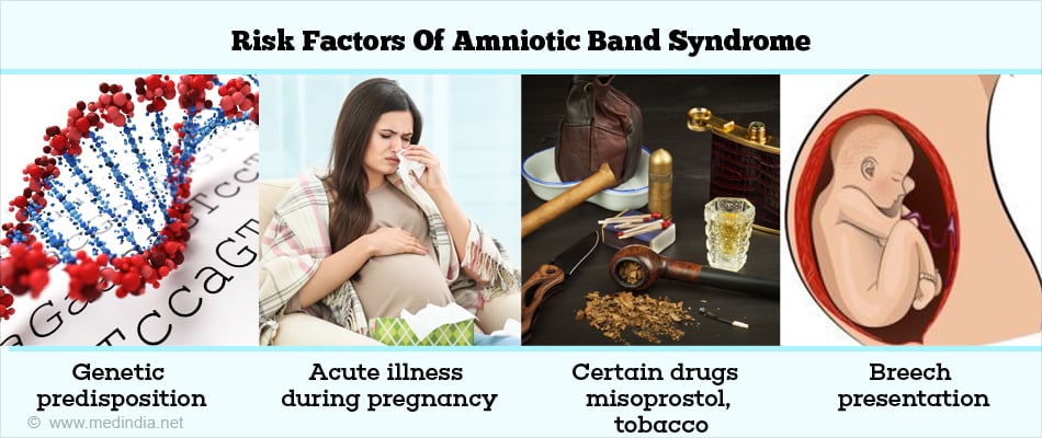
Several factors have been shown to be associated with an increased risk of ABS, but none are conclusively proved. These include the following:
- Hypoxic mechanism due to living at high altitudes
- Family history of Amniotic Band syndrome indicating genetic predisposition
- History of acute illness during pregnancy
- Ingestion of certain drugs such as misoprostol, tobacco
- Diabetes in the mother
- Presence of birth defects such as club foot, cleft lip and palate, and hemangioma (noncancerous growth of blood vessels)
- Non-cephalic presentations of baby like breech position

What are the Symptoms and Signs of Amniotic Band Syndrome?
Amniotic Band syndrome shows varied range of presentation with no two cases similar. Several different patterns of amniotic band syndrome are seen. In general, the more severe forms of amniotic band syndrome occur when the constriction occurs in the first trimester.
The three most common patterns are:
- Serious malformation of the arms and legs
- Limb-body-wall complex characterized by one or more limbs and body wall being affected
- Craniofacial characterized by abnormalities of the head and face and birth defects of the brain and spinal cord (neural tube defects)
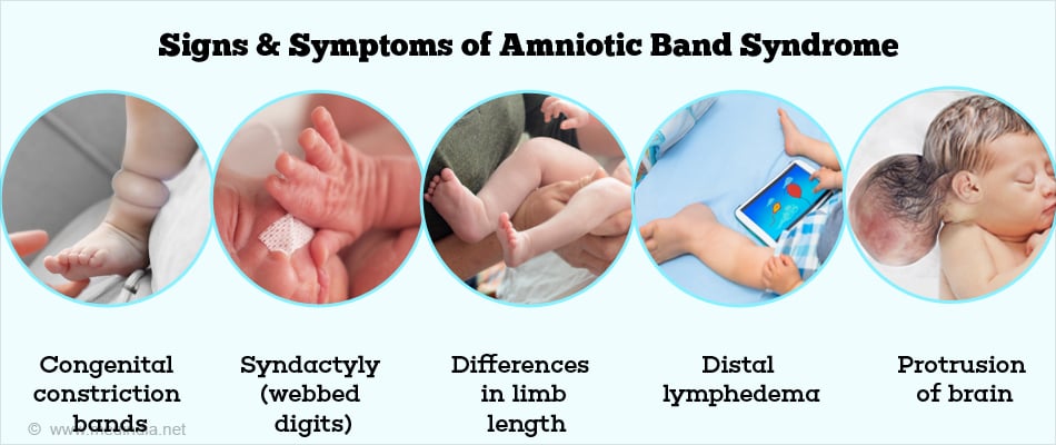
The various clinical features encountered include the following:
- Congenital constriction bands
- Syndactyly (webbed digits)
- Distal ring constrictions
- Nail deformities
- Stunted growth of small bones of fingers and toes
- Differences in limb length
- Distal lymphedema (swelling)
- Limb amputation
- Club foot
- Cleft palate, cleft lip, small eyes (microphthalmia) and other craniofacial defects if band entraps the face
- Neural tube defects such as anencephaly
- Protrusion of brain and meninges through skull defects
- Abdominal wall defects with protrusion of abdominal viscera
- Chest wall defects with protrusion of thoracic structures
- Numerous miscarriages, such as when a band twists around the umbilical cord

How do you Diagnose Amniotic Band Syndrome?
After birth
Amniotic band syndrome is diagnosed typically soon after the baby is born based on characteristic physical findings. Minimal diagnostic criteria consist of the detection of characteristic defects of the arms, legs, fingers, and/or toes, such as ring-like constriction bands or amputation defects.
Diagnostic imaging including x-ray to determine the extent of bands into the deeper tissues and magnetic resonance imaging (MRI) or other scans may be necessary to see how the bands affect blood vessels and nerves.
Before birth
- Occasionally the condition may be diagnosed before birth, either incidentally during routine ultrasound or if suspected, like for women with known risk factors. During fetal ultrasonography, high-frequency sound waves are used to produce an image of the developing fetus.
- Other tests that may raise a suspicion of the diagnosis include amniocentesis where a small amount of amniotic fluid is drawn out and analyzed for increased levels of alpha-fetoprotein.
- Fetal echocardiogram (ultrasound examination of fetal heart) may be done to rule out defective heart development, particularly if banding is seen over the abdominal wall.

One extremely rare inherited disorder called Adams-Oliver syndrome (AOS) has symptoms similar to amniotic band syndrome and can make diagnosis difficult. AOS is characterized by defects of the scalp and abnormalities of the fingers, toes, arms, and/or legs.
Hence accurate diagnosis is important for management of the present pregnancy as well as counseling for future pregnancies.
How do you Treat Amniotic Band Syndrome?
If the bands are not deep and do not cause any symptoms or health problems, no treatment may be required. Treatment is mainly supportive and symptomatic.
Surgery
Some babies need surgery immediately after birth to release the bands if there is concern about damage to blood vessels or nerves and severe swelling (lymphedema) of the limb. In other cases, surgery may be delayed for upto six months of age. Surgery may be done to give the affected part a normal appearance.
There have also been reports of in-utero surgery performed to remove bands that threatened fetal development.
Other treatments:
- Realignment surgery to restore affected bones to better position
- Compression garments to control pressure
- Rehabilitative equipment to help the child cope with the condition
- Prosthetics to replace body parts that are absent or underdeveloped
- Physical and occupational therapy to obtain optimal use of affected limbs
- Oxygen support in babies with underdeveloped lungs

Treatments under Investigation
Studies are ongoing to identify the presence of underlying risk factors such as genetic factors that may increase the chances of occurrence of amniotic band syndrome. These may shed light on the complex mechanisms involved in the development of amniotic band syndrome.


