Decoding of the COVID-19 spike protein structure has enabled a better view of their mechanism of infection in host cells.
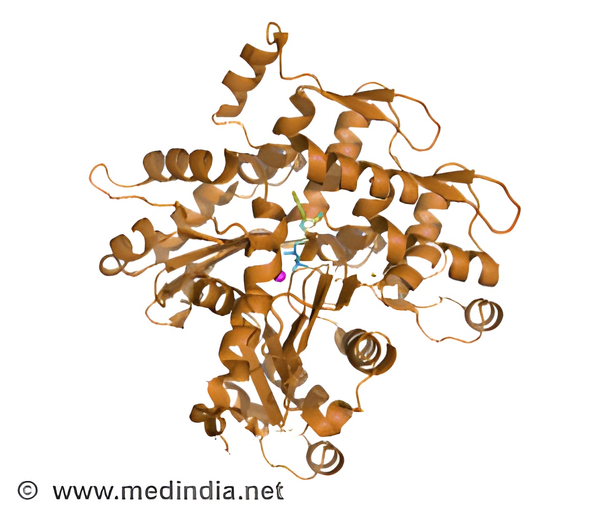
‘Decoding of the COVID-19 spike protein structure has enabled a better view of its mechanism of infection in host cells. This aids in designing better therapeutic drugs and vaccines against the virus.’


There are seven infection causing strains of coronaviruses, among which four cause relatively mild illnesses and the other three - including SARS-CoV-2, the virus that causes COVID-19, being deadly. As the virulence of the virus based to the current pandemic, high enough level of biosafety controls are required in labs for studying the virus. This explains the reason why present study incorporated milder form of the virus — NL63 for further investigation. The virus attaches to humans in a similar fashion to one causing qCOVID-19, thereby causing common cold symptoms. It accounts for 10% of human respiratory disease each year.
The spike protein structure:
The scientists purified NL63's spike proteins and flash-froze whole intact viruses into a glassy state thereby preserving the natural arrangement of their components. Digitally the spike protein copies were extracted to analyse them. The technique had better realistic outcome by skipping the prior formaldehyde fixation of virus.
“The structure we saw had exactly the same structure as it does on the virus surface, free of chemical artifacts. This had not been done before”, says co-author Jing Jin, an expert in the molecular biology of viruses at Vitalant Research Institute in San Francisco.
Advertisement
Thus the study emphasizes on extracting the real essence of the mechanism by which the spike protein binds to receptors on human cells and it’s further activation.
Advertisement

