How do you Diagnose Thymoma and Thymic Carcinoma?
Your physician will first take your medical history, such as age and any potential symptoms, such as myasthenia gravis. When a mass is detected in the chest x-ray, a computed tomography (CT) scan of the chest can distinguish the tumor features and its spread to surrounding regions. Positron emission tomography (PET) helps to distinguish between invasive and noninvasive thymomas. Contrast imaging detects blood vessel invasion of tumors.
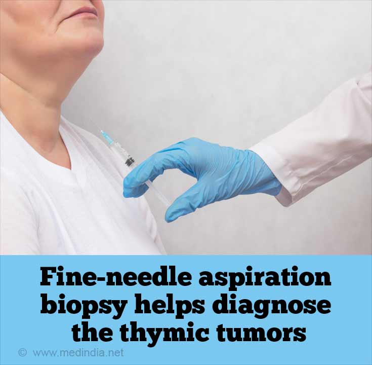
A biopsy (needle core biopsy; fine needle aspiration) provides a small mass of tissue to determine the presence of thymoma at a generalized level. Biopsies may be performed before or after surgery. WHO guidelines for staining cells (histologic classification) help to distinguish thymomas from thymic carcinomas. Antibodies to distinct proteins help to clearly distinguish different types of thymomas (e.g. A, AB, B1). For example, an abundance of lymphocyte cells is observed in types AB, B1, and B2. However, you cannot predict the effectiveness of treatment or recovery based on histologic stains.

Staging (Masaoka-Koga stage) detects the invasiveness of the tumor to determine the type of treatment required. Thymic tumors are classified as:
- Stage 1: Localized tumors covered with a capsule
- Stage II: Initiation of invasion through the capsule (IIa) or into the surrounding fat (IIb)
- Stage III: Tumors invade an adjacent organ, such as the heart and lungs
- Stage IV: Spread of the tumors in the lung or heart membrane (IVa) or invasion of the lymph or blood vessel network
During staging, magnetic resonance imaging (MRI) helps to confirm the presence of associated tumors.
Some of the characteristic diagnostic features of thymoma are:
- A well-defined tumor mass covered with a capsule
- Absence of cancer anywhere else in the body
- Tumor located in the chest cavity (anterior-superior or anterior mediastinal compartment)
- Presence of paraneoplastic conditions, such as myasthenia gravis, red cell aplasia, among others.
How can you Treat Thymoma and Thymic Carcinoma?
Surgery is the primary treatment option for thymoma and thymic carcinoma. Complete resection improves survival in stages I and II tumors. Surgeons and pathologists should communicate well to coordinate the appropriate treatment regimen. When the tumor is encapsulated, it is a localized tumor and should be completely resected. However, with advanced tumor stages (III and IVa), chemotherapy is considered before surgery.
Chemotherapy may be provided post surgery in advanced stages of thymic tumors. Some of the drugs used are cisplatin, doxorubicin, and cyclophosphamide, among others.
External beam radiation therapy following surgery is recommended after complete or partial tumor resection. In most cases, radiation therapy is provided to stages IIb, III, and IVa tumors.
Targeted therapy, e.g. anti-angiogenesis therapy is used to target thymic tumors by blocking the blood supply to the tumor.
Hormone therapy (corticosteroids) may be used to treat thymic tumors. Hormones may block the growth of tumors by inhibiting certain growth-stimulating proteins.
When tumors spread to other parts of the body (stage IVb), the existing treatment regimen cannot cure the tumor. Palliative chemotherapy is the only option in this case.


