- What are Kidney Stones? - (https://www.urologyhealth.org/urologic-conditions/kidney-stones)
- Calcium-Regulating Hormones and the Kidney - (https://www.kidney-international.org/article/S0085-2538(15)34142-9/pdf)
- Diagnosis of Urolithiasis and Rate of Spontaneous Passage During Pregnancy - (https://www.ncbi.nlm.nih.gov/pubmed/22014825)
- Management of Urolithiasis in Pregnancy - (https://www.ncbi.nlm.nih.gov/pmc/articles/PMC3792830/)
- Symptomatic Nephrolithiasis Complicating Pregnancy. - (https://www.ncbi.nlm.nih.gov/pubmed/11042313)
- Renal Stones in Pregnancy - (https://www.ncbi.nlm.nih.gov/pmc/articles/pmc4934980/)
- Kidney Stones - (https://kidshealth.org/en/parents/kidney-stones.html)
- Stones in Pregnancy and Pediatrics - (https://www.ncbi.nlm.nih.gov/pmc/articles/PMC6197562/)
Kidney Stones in Pregnancy
Kidney stones in pregnancy are a challenge both from the point of diagnosis and treatment. They are most common during the first three months of pregnancy with almost 80 to 90% presenting during this period. During this time radiation to the fetus is a hazard and hence doing an X-ray or a CT scan for diagnosis is difficult.
Most of these stones are relatively small and over two-thirds will pass out spontaneously in the urine. Those that do not pass easily, can cause obstruction and pain and can induce premature labor leading to abortion. Giving anesthesia to treat such stones by endoscopic procedures too has similar hazards.
The colicky pain of kidney stones can mimic the pain of appendicitis, placental detachment or diverticulitis hence diagnosis becomes difficult clinically without any investigations. Not diagnosing the condition in time, can lead to complications like obstruction and infection of the kidney and this can create an emergency situation. All this means that immediate care is important when a stone is suspected.(1✔ ✔Trusted Source
Renal Stones in Pregnancy
Go to source)
The risk of kidney stones in pregnancy is estimated to be about 1 in 1500 and is similar to non-pregnant women. However this incidence may be rising due to the rise in global temperature.
Why do Kidney Stones form during Pregnancy?
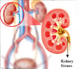
The kidneys are the filtering units of the body. They are two bean shaped organs present just below the rib cage, one on either side of the spinal column. Blood carrying impurities gets filtered after entering the kidneys; this process results in urine formation. The urine drains from the kidney, passes through the ureters and into the urinary bladder. Urine is then voided out through another small tube called the urethra.
The urine is a mixture of waste chemicals and minerals. These chemicals sometimes form small crystals which then fuse together to form kidney stones.(2✔ ✔Trusted Source
What are Kidney Stones?
Go to source)
Kidney stones are medically also called 'Urolithiasis' (Uro-related to urology and lithiasis in Latin means stones). It is a condition where stones are found in the urinary tract, which includes the kidneys, ureters or the urinary bladder. They range from microscopic crystals to stones as large as 3-4 cm.
Women could be more prone to developing kidney stones during pregnancy for the following reasons:
a. Urinary calcium excretion increases during pregnancy. This is due to increased absorption of calcium from intestine, increased re-absorption of calcium from bone and due to the hormonal effects of the parathyroid hormone. Since more calcium reaches the kidneys, it has the possibility of precipitating and forming stones, especially if the woman does not drink enough water.
b. As kidney filtration increases in pregnancy, there is also increased excretion of uric acid, which could increase the chances of uric acid stones.
However, substances that inhibit stone formation are also excreted in the urine to a greater extent during pregnancy. Therefore, the overall risk for kidney stone development is not increased during an otherwise normal pregnancy.(3✔ ✔Trusted Source
Calcium-Regulating Hormones and the Kidney
Go to source)
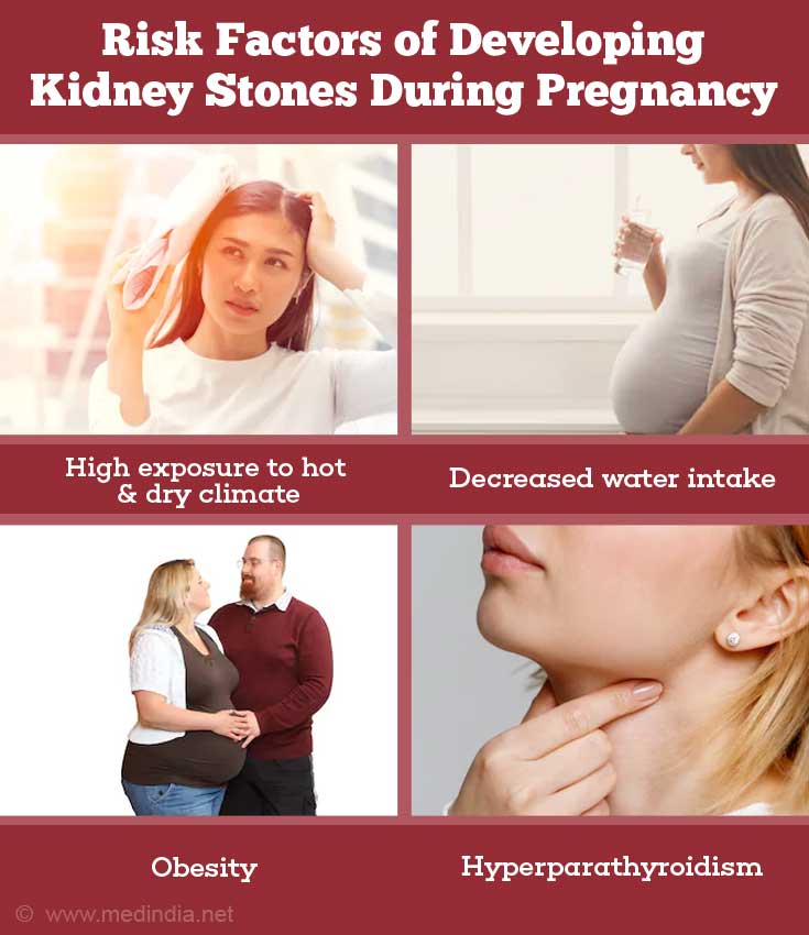
What are the Risk Factors for developing Kidney Stones during Pregnancy?
Risk factors for developing kidney stones during pregnancy include the following:
- Middle-age between 30-50 years
- Continuous exposure to hot and dry climate (like in farm workers and laborers)
- Decreased water intake and subsequent decreased urinary output. This leads to increased saturation of the urine with calcium and other minerals
- Presence of a family history of kidney stones
- Intake of foods rich in calcium, sodium and red meat
- Obesity
- Presence of inflammatory bowel disease (Crohn’s disease and ulcerative colitis)
- Presence of spinal cord disorders
- Hyperparathyroidism
- Presence of birth defects or abnormality in the structure of the kidneys, ureters or urinary bladder
What are the Symptoms and Signs of Kidney Stones?
Generally the symptoms and signs of kidney stones during pregnancy are similar to those caused by kidney stones at other times.
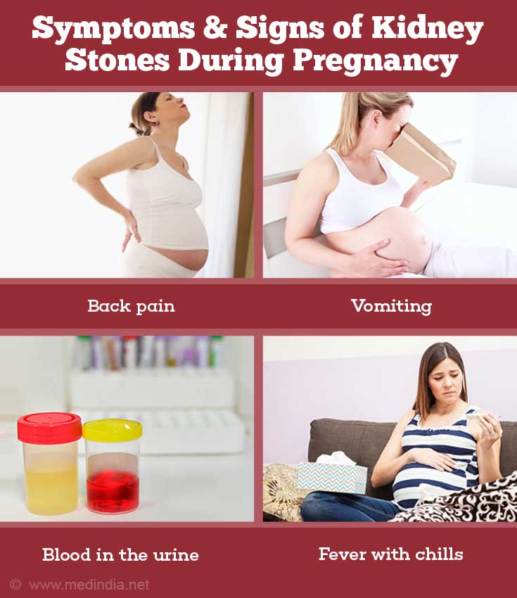
Pain is one of the commonest symptoms of kidney stone and the site of pain depends on the location of the stone. If the stone is in the kidney the pain is usually fixed to the back region below the rib cage. However if the stones move down the ureter, the pain will radiate down towards the side of the body.
- Flank pain or back pain -
- The pain starts suddenly and is severe in intensity. The degree of pain depends on the extent to which it causes obstruction
- Once the stone moves down to the ureter - the pain radiates from the flank to the groin or to the inner side of the genitalia or labia or even go down the thigh
- Lower abdominal pain - The pain should be differentiated from other conditions like appendicitis and placental abruption
- Pain while passing urine especially towards the end - this means the stone has traveled down but still stuck in the lower end of the ureter
- Blood in the urine - maybe frank blood or maybe seen only under microscope
- Nausea, vomiting
- Fever with chills or rigors - indicates possibility of infection
- Occasionally, distension of the abdomen
How do you Diagnose Kidney Stones during Pregnancy?
Investigations used to diagnose kidney stones in pregnancy include the following:(4✔ ✔Trusted Source
Diagnosis of Urolithiasis and Rate of Spontaneous Passage During Pregnancy
Go to source)
Blood and urine tests:
- Urine analysis may reveal blood in the urine, as well as other features calcium or uric acid crystals
- Kidney function tests: Serum urea, serum creatinine are raised and eGFR (estimated glomerular filtration rate) is reduced if there is damage to kidneys
- Urine culture and sensitivity identifies the organisms causing urine infection, and the antibiotic to which they are sensitive to. Infections may occur following obstruction to the urinary flow caused by the kidney stones
- Complete blood picture and CRP (C reactive protein) detect inflammation or infection in the body
- Changes in serum electrolytes like increased potassium and decreased serum bicarbonate levels indicate kidney tubular acidosis
- Serum calcium may be elevated in hyperparathyroidism
- Serum uric acid may be elevated, predisposing to kidney stones
Imaging studies in Pregnancy
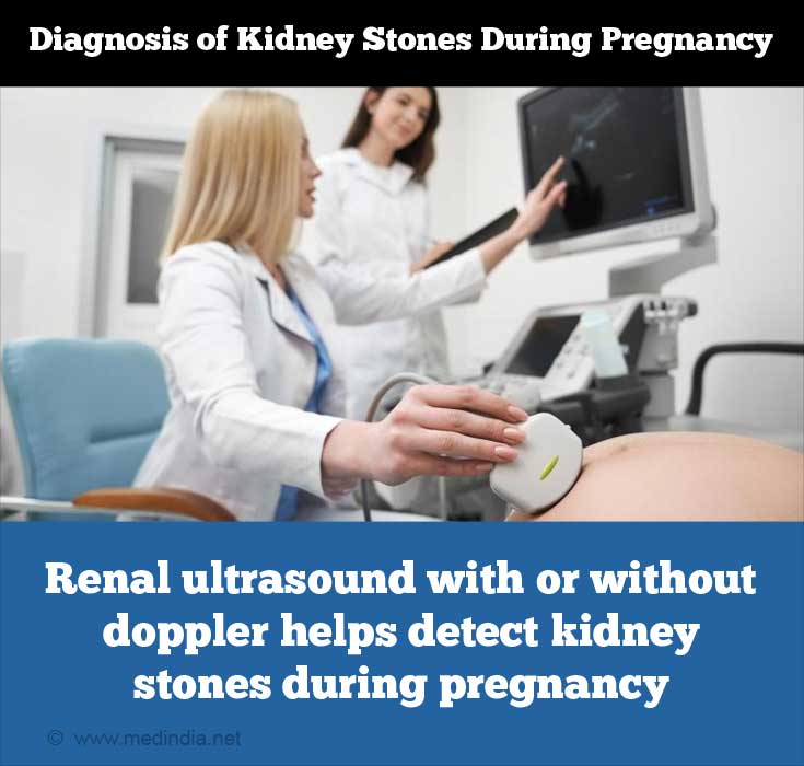
a. Renal Ultrasound: Kidney ultrasound with or without doppler studies is the primary imaging investigation. It is cheap, easily available, can be done without pain and radiation exposure to the fetus, and without injecting any allergenic contrast material into the patients. Doppler studies help to evaluate the kidney blood vessels, which can give clues to the presence of an obstruction in the urinary tract. The disadvantage of using ultrasound is that it cannot identify some types of kidney stones, and cannot differentiate enlarged kidneys due to normal pregnancy or obstruction from a stone.(5✔ ✔Trusted Source
Management of Urolithiasis in Pregnancy
Go to source)
For stones in the lower end of the ureter, which cannot be visualised on a regular ultrasound, a trans-vaginal ultrasound (the ultrasound probe is inserted through the vagina) may be helpful
b. X-Rays and CT-scans - Plain CT scan to diagnose stones in the urinary tract is currently the gold standard test and is routinely performed in all such cases. However it is best avoided during pregnancy due to the effects of ionizing radiation on the unborn fetus. The risks to the unborn baby of radiation are more during the first six months of pregnancy when there are rapid divisions of cells. Hence these tests are best avoided. If they are done the risk should be explained to the parents.
Many experts have the view that the risks of cancer or genetic defects are minimum if the radiation exposure is less 50 mGy (mGy - is unit of radiation and stands for milligray. One milligray is equal to one thousandth of a gray and the gray is defined as the absorption of one joule of ionizing radiation by one kilogram of matter, e.g. human tissue)
An X-ray of the abdomen produces an exposure of 4.2 mGy whereas a CT scan causes exposure of 49 mGy.
If x-ray or CT is planned for diagnosis of the stone, the radiologist will reduce the fetal radiation exposure to the minimum by keeping the exposure time or number of x-rays to the least required. They will also use lead shields to protect the maternal pelvis.(6✔ ✔Trusted Source
Symptomatic Nephrolithiasis Complicating Pregnancy.
Go to source)
c. Role of Limited IVU (intravenous urogram) - If ultrasound has been unsuccessful in identifying the stone but the presence of stones is strongly suspected and CT scan is not available - a limited IVU without using full dose of radiation can be done. An x-ray of the kidney, ureters and bladder are taken first, then contrast material is injected and another x-ray is taken after 30 minutes.
c. Magnetic Resonance Imaging (MRI): MRI does not use radiation or contrast materials, hence it is safe but is normally not advised in the first trimester. Though stones may not be identified directly, MRI can identify the point of obstruction.
What are the Indications for Surgical Intervention in Pregnant Women with Kidney Stones?
Surgical intervention is advised when conservative management fails. Indications are:
- Stones causing obstruction of the urinary tract leading to pyelonephritis or sepsis of the kidneys
- Obstruction in a patient having only one kidney
- Acute kidney failure
- Severe pain
- Premature labor triggered by kidney pain and not settling with labor suppressant drugs
What is the Treatment of Kidney stones in Pregnancy?
The goal of treatment should be to minimize the symptoms in the mother, to prevent kidney damage and sepsis, and to minimise risk to the foetus.(7✔ ✔Trusted Source
Kidney Stones
Go to source)
Hospital admission is advised.
Conservative management should always be provided first. This includes:
- Bed rest
- IV fluids
- Pain killers/analgesics
- Antibiotics
- Anti-nausea/vomiting medication
With this, approximately 70-80% stones will spontaneously pass out or the symptoms would settle down sufficiently for
The surgical procedures will require anesthesia, and the procedures generally advocated are the following:
- Ureteroscopy: A small diameter tubular telescope is passed into the ureters via the urethra and the urinary bladder. This is both a diagnostic as well as a therapeutic procedure. Stones can be easily identified and removed at the same time. Advantages include the use of single surgical procedure for the diagnosis as well as the treatment, the quick resolution of symptoms and avoidance of stent or nephrostomy tube-related complications. Laser is the preferred source to break the stone if required.
- Ureteral stent placement: A hollow, soft silastic tube is placed into the ureter under cystoscopy guidance. This helps to temporarily relieve the obstruction and the drain out the urine. The stents have to be replaced every 2 to 3 months. Definitive treatment is then delayed until after delivery.
- Percutaneous nephrostomy: A small tube is placed into the kidney through the skin. This helps with quick decompression of the kidney, rapid control of pain and resolution of the infected enlarged kidney.
- Open surgery or ESWL (shockwave lithotripsy) is not routinely advised in pregnant women.
How can Kidney Stone Complications be Prevented in Pregnancy?
Kidney stone complications during pregnancy can be prevented by:(8✔ ✔Trusted Source
Stones in Pregnancy and Pediatrics
Go to source)
- Increasing fluid intake (around 2 litres/day)
- Reducing salt intake
- Reducing the consumption of foods like chicken, fish, red meat, beef, peanuts, chocolate, nuts, green leafy vegetables, coffee, tea and beetroot
- Avoiding excessive calcium intake. The daily recommended dose of calcium should not exceed 1000-1200 mg/day. Conversely, very low calcium diets are also not advised
Health tips
- High citrate content of lemon juice and
citrus fruits inhibits stone formation - Limit intake of high salt and sugary food
- Limit alcohol intake as it can cause uric acid stones
- Eating more fruits and vegetables will reduce urine acidity and stone formation.
- Women who have kidney stones or are at high risk for kidney stones may be advised to undergo diagnostic tests and kidney stone removal before planning pregnancy


