- J P Fisher. Pterygium - (http://emedicine.medscape.com/article/1192527-overview)
- Boyd E. What is pinguecula and pterygium? - (https://www.aao.org/eye-health/diseases/pinguecula-pterygium)
What is Pinguecula?
Pinguecula (pronounced as “pin-GWEK-yoo-lah”, plural is pingueculae) is a common lesion of the eye. It is a yellow, or yellowish-white, opaque, soft, harmless, small growth found on the sclera (white part of the eye), in between both the eyelids. It is derived from the Latin word “Pinguis” which means “Fat”,because of its appearance to fat tissue; however it does not contain any fat.
Anatomy of the Eye
The external eye is made up of eyelids and eyelashes. The white part of the eye is called the sclera; it is a tough opaque layer that protects the inner structures of the eye. The conjunctiva is a thin transparent film like layer that covers the sclera (bulbar conjunctiva) and extends to the inner side of the eye lids (palpebral conjunctiva). This layer helps to keep the eyes moist by secreting mucous and a small amount of tears. The cornea is the clear, convex shaped thin membrane like structure covering the iris (the coloured part of the eye) and the pupil (the black opening in the centre of iris). The cornea directs the light into the eye and focuses it on the retina. The junction of the cornea and the sclera is called the limbus.

Location of Pingueculae
Pingueculae are located on the bulbar conjunctiva and can be of varying sizes. They are oval or round but can be triangular with the base at the limbus. They are usually horizontally placed rather than vertical. Pingueculae are commonly bilateral, but can be present in only one eye too. It is They are frequently present on the sclera towards the side of the nose (nasal side), but can sometimes be seen on the temporal side too (the sclera towards the ear).
Mechanism of Pinguecula Formation
It is formed due to thinning of the top layer of the conjunctiva, degeneration of the inner layers of the conjunctiva and deposition of protein or fat. Sometimes it can undergo calcification.
A pinguecula if formed as a result of elastotic degeneration of the superficial layers of the conjunctiva. There is deposition of hyaline material, and occasionally small areas of calcium deposition may be seen.
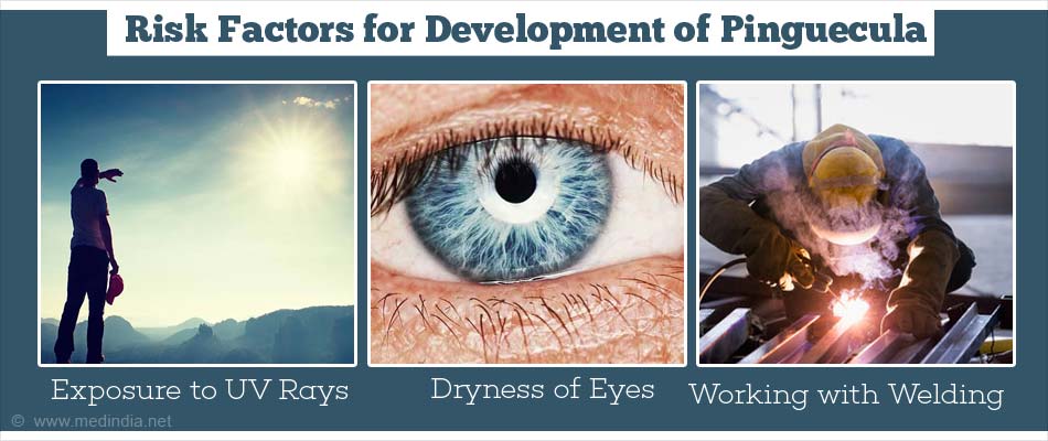
What are the Risk Factors for Development of Pinguecula?
- Pinguecula is more common in individuals over 40 years of age, but is also seen in younger individuals who are constantly exposed to sunlight, windy or dusty environments. This is the reason it is known as Farmer’s eye.
- Constant exposure to ultraviolet radiation (both A and B rays) is an important risk factor in people of all ages including children.
- People near the equator are more prone to develop pingueculae due to exposure to strong sunlight. It has been suggested that pingueculae are commonly found on the nasal side of eye due to reflection of light from the nose onto the nasal side conjunctiva.
- Males are affected more than females probably because of more ultraviolet exposure.
- Patients already having dryness of eyes due to a pre-existing cause and contact lens wearers may be at risk to develop pingueculae
- People working with welding arc can develop pingueculae due to exposure to strong light.
- Smoke
- Diabetes Mellitus
- Gaucher’s disease
What are the Signs and Symptoms of Pinguecula?
Pingueculae do not cause symptoms in the majority of people.
As a pinguecula is a raised bump, the natural tear film spreads unevenly over the surface of the eye, thus causing a break in the tear film. This leads to dryness of eyes, burning sensation, itching, constant rubbing of the eyes due to foreign body sensation, blurry vision and stinging.
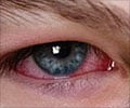
Sometimes, the pinguecula can become inflamed leading to swelling and redness. This is called pingueculitis and can be painful. A large pinguecula can look cosmetically unsightly.
How is Pinguecula Diagnosed?
It is diagnosed clinically. A slit lamp examination gives detailed information by providing a magnified view of the external eye structures. Here a light is shone into the eye and the doctor looks through a microscope from the other end. In atypical cases, biopsy of the growth is taken and a pathologist studies the specimen under a microscope to confirm the diagnosis.
How is Pinguecula Treated?
The majority of patients do not need treatment and simple reassurance about the harmless nature of the condition is sufficient.
Medical management:
If the symptoms are severe then treatment is needed.
- Lubricating eye drops during the day and ointment during the night can be used if there is dryness or foreign body sensation.
- Scleral contact lenses are another option – these are used to cover the pinguecula, protecting it from further exposure to UV rays and from dryness.

- If patient has pingueculitis –along with cold compresses low potency steroid eye drops like loteprednol, rimexolone or fluorometholone can be used. Non-steroidal anti-inflammatory eye drops like indomethacin can be used too. Topical steroids and NSAIDs will reduce the inflammation. Long term use of steroid eye drops can cause a rise in intraocular pressure and is not recommended for a pinguecula.
Surgical management:
Surgical removal of the pinguecula is only considered if the symptoms are severe or recurrent, if it interferes with person wearing contact lenses, if the patient finds blinking difficult or if it is cosmetically bothersome.
There have been a few patients whose pinguecula was treated with argon laser photocoagulation; further research is awaited here.
What are the Complications of Pinguecula?
- Pinguecula can develop into a pterygium.
- The cornea next to the pinguecula can dry out and this leads to corneal thinning. This is called dellen.
- Pinguecula may also cause chronic actinic keratopathy.
How can you Prevent Pinguecula Formation or from getting Worse?
- Avoid sunlight and UV exposure by wearing a wide brim hat.
- Eyes can be protected from irritants like sunlight, UV radiation, dust, wind etc. by wearing good quality wraparound sun glasses (glasses with a frame which wraps around the eyes more than the normal frame)
- In regions where it snows, it is important to still wear sunglasses in winter, as the snow reflects about 80% of UV rays.
- Use eye lubricants to moisturize the eyes

Health tips:
- Cold compresses can help settle pingueculitis.
- Do not use steroid eye drops even for a short duration, unless it is prescribed by a doctor, as steroid drops can cause unwanted side effects.
- Wear wide brim hat and sunglasses even on cloudy days, as UV rays can penetrate cloud cover.
- People working outside with sand, concrete, water, snow etc. should also use sunglasses as these surfaces reflect sunlight.


