- Retinitis Pigmentosa Treatment - (https://www.aao.org/salud-ocular/consejos/retinitis-pigmentosa-treatment)
- Treatment for Retinitis Pigmentosa Reported - (https://nei.nih.gov/news/pressreleases/rppressrelease)
- Retinitis Pigmentosa and Retinal - (https://www.asrs.org/patients/retinal-diseases/8/retinitis-pigmentosa-and-retinal-prosthesis)
- How Is Retinitis Pigmentosa Diagnosed? - (http://www.visionaware.org/info/your-eye-condition/retinitis-pigmentosa/how-is-retinitis-pigmentosa-diagnosed/125)
- Retinal diseases - (http://www.mayoclinic.org/diseases-conditions/retinal-diseases/care-at-mayo-clinic/tests-diagnosis/con-20036725)
- Facts About Retinitis Pigmentosa - (https://nei.nih.gov/health/pigmentosa/pigmentosa_facts)
- About Retinitis Pigmentosa - (http://www.blindness.org/retinitis-pigmentosa)
- Information About Retinitis Pigmentosa - (http://eyewiki.aao.org/retinitis_pigmentosa)
- Retinitis pigmentosa - (https://en.wikipedia.org/wiki/retinitis_pigmentosa#causes)
What is Retinitis Pigmentosa?
Retinitis pigmentosa is an inherited degenerative disorder which affects the retina’s ability to respond to light. It results in progressive loss of vision, eventually leading to blindness. It involves both eyes.
There is no cure for retinitis pigmentosa. Early intervention can help retain the vision and make the most of it.
What is Retina?
Retina is a light sensitive layer lying at the back of eye. Light first focuses on the eye lens and then passes through the pupil and falls on the retina. Light rays that fall on the retina are converted into neural signals which are then sent to the brain through the optic nerve. Thereafter, the brain interprets and sends the signal for visual recognition.
Retina is made up of photo-receptor cells. These cells perform the function of detecting color and light intensity. These are of two types – rods and cones. Rods lie on the periphery of the retina and support black and white vision. They focus on distant vision and dim light. Cones are centrally located in the retina and support colored vision and near sighted vision.
In retinitis pigmentosa, these cells get degenerated and stop working leading to loss of vision. Disease starts with death or necrosis of rods; eventually cones are affected leading to total blindness. Side vision is impacted, the most owing to the rods, which makes it hard to see in dim light. Early intervention helps retain the cones so that near sighted vision to a large extent can be preserved.
What are the Causes of Retinitis Pigmentosa?
Retinitis pigmentosa (RP) is genetically inherited. It is passed from the parent to the child. Let’s understand how.
Each cell of the body contains 23 pair of chromosomes. Out of these 23, 22 pair of chromosomes carry non sex traits and 1 pair of chromosome carries sex traits. Males have one X and one Y chromosome (XY) whereas females have two X chromosomes (XX). During conception, when the sperm fertilizes the egg, one copy of each chromosome is passed onto the fetus. Retina pigmentosa gets inherited when the following combinations are formed during conception:
- Autosomal recessive – In this case both, mother and father are carriers of the mutant gene but are not suffering from RP disorder. Retinitis pigmentosa occurs when one copy of the same mutated gene is passed on by the parents to the individual. In a pair of genes, if one is normal and other is mutated, the individual will be a carrier for disease and will not have the dominant disease.
- Autosomal dominant – In this case, either the mother or the father is suffering from this disorder and has one copy of the dominant gene that is passed on to the child.
- X- Linked – If the mother’s sex linked gene (X) is mutated and passed on to the child, it results in disease inheritance. Males have only one X chromosome. If and when a mutant copy of X is passed on, the child will develop the disease. A male with an X-linked disorder passes the disease gene to daughters and not to sons, since the X chromosome of the male goes to the daughter and the Y chromosome goes to the son. So all his daughters will definitely be carriers of the disease. Males are more often and severely affected than females. Females have two X chromosomes. If the female child receives one of the normal copy of X and one mutant copy of X, then the child becomes a carrier and remains asymptomatic. If the female child receives both the mutant copies, then the child inherits the disease.
What are the Symptoms and Signs of Retinitis Pigmentosa?
Retina pigmentosa:
- Begins with difficulty seeing in dim light - like at night or at dusk. This is also known as night blindness. It takes a maximum of 20 minutes for an eye to get adapted to dim light. But in case of retinitis pigmentosa, the eye takes much longer or the eye will not get adapted to the dim light at all, resulting in night blindness.
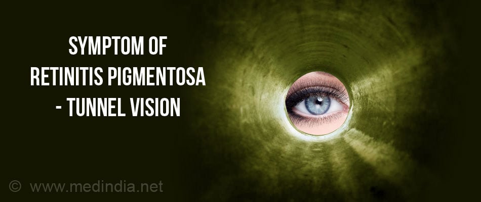
- This is followed by loss of side vision, wherein the individual will not be able to see things on the side, above or below. He will only be able to look straight. The rate of progression varies from person to person. Symptoms might appear as early as 10 years. By the early 40’s, most people with retinitis pigmentosa have only restricted vision, with very narrow visual field. They can only look straight through that field, as if looking through a tunnel. This is called “tunnel vision”.

- As the disease progresses, visual clarity and visual acuity decrease. By the time the person is in the 50s, near sighted vision also becomes difficult, since centrally located cones also start getting affected and a magnifier is required for reading and recognizing. People with retinitis pigmentosa find it difficult to go out in bright sunny light, as adaptation from bright sunny light and glare to the one inside an enclosed area becomes a problem.
How do you Diagnose Retinitis Pigmentosa?
- Comprehensive Eye Examination - Doctor examines the individual’s eye with an ophthalmoscope. Healthy retina appear orange red in color, but when an individual has retinitis pigmentosa, black or brown pigmentation appears on the orange surface of the retina. This is quite an early sign.
- Visual Field Testing - This helps in detecting the extent of the damage. It helps determine how much side vision is left. It is done by a non-computerized visual field test, such as the Goldmann Perimeter Exam or a computerized one, such as the Humphrey Field Analyzer.
- The Goldmann Perimeter Exam - In this, one eye is covered and the individual is asked to focus straight with the other eye. Doctor passes light from beyond the side of the visual field into the center to find out the location at which the patient can see the light. Doctor analyzes individual’s responses to build a map of the visual field.
- The Humphrey Field Analyzer - In this, one eye is covered and the individual is asked to focus straight with the other eye. Doctor flashes white light of varying intensities and color through the individual’s visual field. The doctor analyzes the individual’s responses to various light colors and intensities to build a map of the visual field.
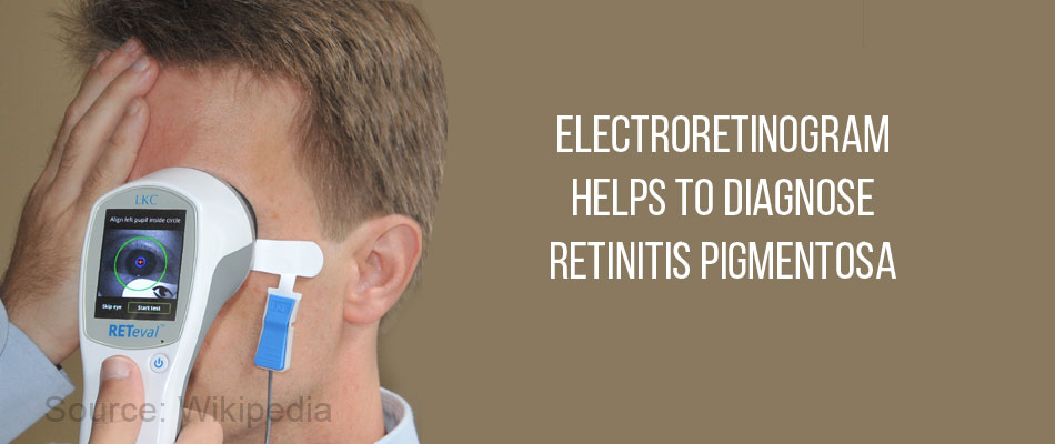
- Electrophysiological Testing - Electroretinogram (ERG) measures the response to flashes of light, via electrodes placed on the surface of eye. It determines the extent of damage to the visual field. Periodic ERG examinations are necessary to track the progression of disease. The ERG measures the electrical activity of rods and cones in response to the light.

- Dark Adaptation Test - It measures the visual adaptability of the eyes in response to dim light. It is very helpful in detecting early cases of rod degeneration.
- Electrooculogram (EOG) - It measures the resting electrical potential between the cornea and the retina. It is a very sensitive test for detection of photo-receptor function and not usually recommended.
- Fluorescein angiography (FA) - A special dye, fluorescein, is injected within the eye that highlights the blood vessels at the back of the eye.
- Optical coherence tomography (OCT) detects the structural changes within the retina resulting from RP. It captures cross-sectional images of the retina.
- Genetic testing - A sample of the patient’s DNA (deoxyribonucleic acid) is taken to check the progression of their particular form of the disease. They can also enroll themselves into a patient registry to take part in clinical trials.
What is the Treatment for Retinitis Pigmentosa?
Currently, there is no cure for retinitis pigmentosa. However, The Foundation Fighting Blindness and the National Eye Institute in the USA have jointly recommended that adults with RP should take a daily dose of vitamin A palmitate supplement and avoid a high dose of vitamin E, as recommended by their doctor. This will help in prolonging their vision.

Once the rods and cones are damaged, the only way of restoring vision is by transplant or by bypassing them, thereby altering the physiology of the eye. Photo-receptor cell transplantation is still under study.
Argus II (Second Sight Medical Products, Inc, Sylmar, CA) is a medical implant certified by the Federal Drug Association (FDA), USA. It consists of a camera and processing device attached to an individual’s glasses. A microchip is implanted in the individual’s retina which is the processing device. The camera captures the information which is transferred to microchip which further stimulates the remaining retinal cell and assists in vision. Another implant, Alpha IMS retinal implant (Retina Implant AG, Reutlingen, Germany), CE approved has also been recommended.
What are the Complications of Retinitis Pigmentosa?
Cystoid macular edema is a complication of retinitis pigmentosa which results in build up of fluid in the macula, the central portion of the retina. This further results in decreasing the near sighted vision since the central visual field starts getting affected. Doctors prescribe medicines to clear out this fluid build up which provides symptomatic relief.


