- SPECT scan - (http://www.mayoclinic.org/tests-procedures/spect-scan/basics/definition/prc-20020674)
- Single-photon emission computed tomography - (http://en.wikipedia.org/wiki/single-photon_emission_computed_tomography)
What is SPECT?
Single Photon Emission Computed Tomography (SPECT) scan uses computed tomography technique to trace the movement of a radioactive material injected into the body. It tracks the blood flow into tissues and organs and also the function of some of the internal organs. It is categorized as a nuclear imaging test where the radioactive substance emits gamma rays, which are in turn detected by a computerized machine. The machine then creates 3-dimensional images based on the readings.
The radioisotopes that are usually used in SPECT include iodine-123, technetium-99, thallium-201, xenon-133 and fluorine-18. These radioisotopes are combined with other natural chemicals like glucose or water to be injected into the body. They travel safely through the blood and are detected by the scanner.
SPECT is different from Positron Emission Tomography in terms of the flow of the tracer. In PET, the tracer is absorbed by the surrounding tissues, thus showing only those areas where the tracer has accumulated. The tracer in the SPECT scan is not absorbed by the tissues and remains in the blood, giving a clear picture about how blood flows into the heart or what areas of the brain are more active.
Why is SPECT done?
SPECT scan is recommended for patients who have heart problems, brain disorders or bone disorders. The results of a SPECT scan helps in understanding the disorders associated with the blood flow and supply to the parts of heart, brain and bone. SPECT is used for:
Diagnosis of brain disorders:
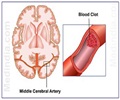
- Identifying blocked blood vessels in the brain, which could result in transient ischemic attacks and stroke
- Epilepsy and Seizures
- Dementia
- Trauma to brain due to head injury
Diagnosis of heart problems:
- Block in the coronary arteries: Parts of the heart muscle supplied by the coronary artery may be blocked and the impaired blood flow is shown.
- Heart contractions: SPECT can be used to check complete draining of blood from the heart chambers when the heart contracts.
Diagnosis and tracking bone disorders:
- Hidden bone fractures
- Diagnose and track progression of cancers that spread to the bones
How is SPECT different from CT, MRI and PET?
The computed tomography (CT) and Magnetic Resonance Imaging (MRI) techniques image the structures of the body. The location and shape of a lesion or a tumor can be understood through a CT or MRI scan.
Positron Emission Tomography (PET) scan is a nuclear imaging technique that uses a radioactive tracer combined with the natural chemicals; it is taken up by tissues.
SPECT scan is a nuclear imaging technique that uses radioactive tracer along with the three-dimensional CT scan images. In SPECT scan, the tracer is meant to remain in the blood without getting absorbed into the tissues. Thus, the blood flow and supply can be better understood with SPECT scan. It also gives high resolution images of the damaged tissues that lack blood supply.
How should a person prepare for a SPECT imaging test?
Discussion with the doctor about your medical history, allergies and so on are important before going for a SPECT scan. Information regarding the procedure and what to expect will be given by the doctor.
Tell the doctor about:
- If you are pregnant or if there is a possibility of being pregnant
- If you are breastfeeding
- Any medical conditions like diabetes or heart disease
- Any medications and supplements taken
- Any allergies present
Things to take care:
- Do not consume tobacco, alcohol or caffeine for at least a day before the SPECT scan
- Do not eat for about 2-3 hours before SPECT scan
When you go in for the SPECT scan:
- Do not wear any metal things in your body, like body piercings, zippers or jewelry
- Empty your bladder
After the scan, the patient may need to avoid going near infants or pregnant women. This is due to the small amount of radioactive tracer that might be present for a while. It is best to drink a lot of water and fluids to flush it out as fast as possible.


