About
A comprehensive eye exam can take an hour or more, depending on the doctor and the number and complexity of tests required to fully evaluate vision and the health of the patient’s eyes.
The list of eye and vision tests that one is likely to encounter during a routine eye examination is as follows:
- Visual acuity
- Retinoscopy
- Autorefraction
- Extraocular movements
- Cover test
- Visual Fields
- Pupillary reaction
- Color vision test
- Slit lamp examination
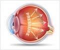
- Intraocular pressure
- Pupil dilatation and retinal examination
Visual Acuity
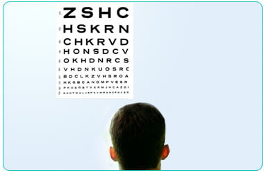
A visual acuity test is a measure of the sharpness and clarity of one’s vision. He will be asked to read letters on a vision chart which is placed at 6 meters or 20 feet. The last line that can be read determines the visual acuity. Visual acuity of 6/6 or 20/20 means the vision is normal.
Retinoscopy
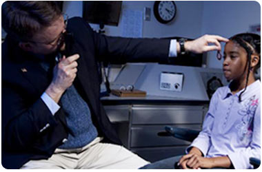
Retinoscopy is a test that gives your eye doctor an idea about the refractive error. It is usually performed after testing for visual acuity. Retinoscopy is performed in a dark room. You will be given a large target to fixate on. The eye doctor will shine a light at your eye and flip lenses in front on your eye. Based on the way the light reflects from your eye, your doctor is able to give your eye glass prescription.
Autorefraction
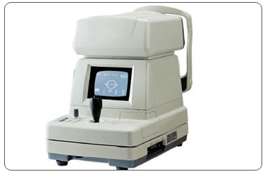
An autorefractor helps detect the lens power required to accurately focus light on your retina. It takes a few seconds and requires you to put your chin on the chin rest and look towards the picture seen straight ahead in the machine without blinking for a few seconds to get an accurate reading. It is a time saving procedure compared to the manual refraction taking only a few seconds.
Extraocular Movements
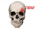
This is a test done to see the movements of the eye in all the directions of gaze. The patient is asked to track a pen or the examiner’s finger which is moved in the patient’s visual field. Any restrictions or weakness in eye movements is picked up which may be due to fractures of the orbital bones, nerve palsies or myopathies.
Cover Test
The cover test is used to detect squinting eyes. The eye doctor covers one eye while you fixate at a distant or near object. While the eye is uncovered the movement of the eye is noticed to interpret the type of squint the patient is suffering from or subtle binocular vision problems causing eye strain.
Visual Field Test
A confrontation visual field is a test done by the eye doctor to know the patient’s uniocular field of view. The patient is made to sit in front of the doctor at about 2 arms length distance. The doctor covers his right eye to check the patient’s right eye field. He/ she brings his/her finger or a red object from the periphery to centre in all the directions 3600from the center. All this while the patient has to look at the examiner’s left eye. The extent of the patient’s left eye field can be similarly tested by closing the doctor’s left eye.
Pupillary reaction
It is done by shining a torch light on the patient’s eye to see his pupillary reaction. The pupillary reaction acts as a window to the health of the nerves that control its function. Abnormal pupillary reaction reveals neurological problems which need further evaluation. It also reveals the health of the optic nerve.
Color Vision Test
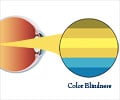
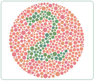
It is ascreening test done in routine eye examination to identify color vision defects and color blind individuals. The most commonly performed test to detect red green color deficiency is Ishihara test plates. It consists of 24 plates with numbers and patterns of different color dots hidden in a background of different colored dots. People with normal color vision can easily identify the number or pattern while those with color defect wrongly identify numbers. There are various other tests to detect color vision like D-15 test, lantern test and holmgreen wool test.
Slit Lamp Examination
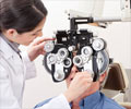
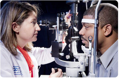
The slit lamp is an instrument used to examine the health of the front portion of the eye. It is called a slit lamp because it uses a slit beam of light to help visualize the ocular structures in a magnified cross sectional view.
You are required to place your chin on the chin rest of the slit lamp machine while the doctor examines your eyes through the oculus of the slit lamp. The front portion of the eye i.e. the lids, conjunctiva, cornea, anterior segment of the eye is visualized in detail. A high powered convex lens is then used to visualize the retina and the optic nerve head.
Eye diseases like corneal ulcers, cataract, infections, diabetic retinopathy, optic nerve thinning, etc can be detected on the slit lamp.
Intraocular Pressure
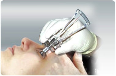
Schiotz tonometer: Measures the eye pressure by virtue of the needle reading on a scale while the tonometer is placed on the black part of your eye. The eye is first anesthetized and the patient is made to lie down. The instrument is then placed over the eye and the needle shows a deflection which is read from a chart for the intraocular pressure.
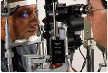
Applanation tonometer: This is a small measuring device attached to the slit lamp. Eye is anesthetized and stained with a yellow dye. The applanation device is then gently touched on the black part of the eye to measure the eye pressure under blue light.
Non contact tonometer: It measures the eye pressure by a puff of air on your eye. There is no contact with the eye and is a painless procedure. Based on the eye’s resistance to the puff of air, the intraocular pressure is measured by the machine.
Schiotz’s tonometer is the means of screening and gives a rough estimate of the eye pressure. Applanation tonometer is a gold standard for accurate eye pressure measurement while the non contact tonometer gives a fairly accurate result with the advantage that it does not touch the eye.
Pupil Dilatation and Retinal Examination
The interior of the eye can be visualized better by dilating the pupil with dilating eye drops. These drops are instilled in the patient’s eyes and he/ she is asked to keep their eyes shut. These drops are put 2/3 times at intervals of 15 mins. This process of dilatation takes around 20 to 30 mins. After which the back of the eye can be examined with the help of a direct or indirect ophthalmoscope.
Ocular conditions like diabetic retinopathy, retinal detachment, retinitis pigmentosa, glaucomatous optic atrophy and many more conditions can be detected by this test.

