Physical Breast Examination
A physical examination or the
In case of fibroadenosis it would be ideal to do this a week after the menstrual periods as this would avoid the discomfort of examining tender breasts. During the exam the patient is expected to take off any necklace and clothes and to wear a gown. Usually a female nurse would accompany the doctor to keep her at ease. The doctor will then talk to the patient to get information regarding her medical history and risk factors after which an examination of the breasts, underarm and collarbone will be carried out to detect signs of injury or infection, changes in breast size, color or texture.
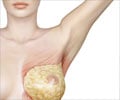
Each breast will be palpated to check for lumps and the nipple gently pressed to detect any discharge.
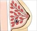
Once the lump is detected the following information about the lump will be noted -
- Precise location of the lump
- How the lump was initially observed and for how long it has been present
- Accompanying nipple discharge, if any
- Change of size / waxing and waning of lump
The "classic" characteristics of cancerous lesions described below helps to identify it from other palpable lumps.
- Single lesion
- Hard and immovable
- Have Irregular borders
- Size > or =2 cm
Once the lumps are detected further tests may be required -
- Chest X-ray
- Mammography,
- Breast ultrasound,
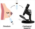
- Fine needle aspiration (FNA), and/ or core needle biopsy


