Diagnosis Of The Carcinoid Syndrome And Carcinoid Tumors
When all the symptoms are present in a patient, a diagnosis of carcinoid syndrome is easily made. Riased urinary 5-HIAA in a patient with a carcinoid tumor is the hallmark for the diagnosis.
Cases that present with one or more of the features are to be viewed with suspicion. A consultation with your doctor should never be delayed.
I. Examination of urine
Most often, the doctor confirms his diagnosis by measuring the levels of certain chemicals in the patient’s urine. Carcinoids produce a number of biologically active substances most which get metabolised. These get excreted by urine.
Excretion of urinary 5-HIAA in a patient with a carcinoid tumor is a diagnostic hallmark 5-HIAA stands for 5-Hydroxyindoleacetic acid. This is a metabolite of the chemical serotonin that gets secreted by carcinoid tumors. Measurement of serotonin or its metabolites in the patient’s urine or plasma gives additional information. Attacks of flushing in a patient accompanied by normal urinary excretion of 5-HIAA it raises the suspicion of diagnoses other than a carcinoid syndrome; for example ethanol ingestion or post menopausal state.
Causes for a false test result
A number of factors interfere with test results. The patient should refrain himself from eating foods like bananas, tomatoes, pineapples, plums, avocados, eggplants, and walnuts. These are rich in serotonin. Cough syrups, muscle relaxants, anti psychotic medications lead to false results. Hence always mention to your doctor about these medications.
II. Imaging techniques to locate the tumor, measure its extent
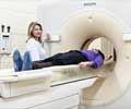
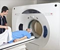
CT scan: In computed tomography (also called computerized tomography, or computerized axial tomography) a series of detailed pictures of areas inside the body are taken from different angles.
MRI scan: MRI stands for Nuclear Magnetic Resonance Imaging. This is a non invasive technique that doesn’t use ionizing radiations unlike a CT scan. Instead strong magnetic fields are used.
Radionuclide scanning: More than 90% of carcinoid tumors can be located using this technique. The principle used is that carcinoids exhibit receptors for the hormone serotonin. A radioactive form of serotonin is injected into the blood and radionuclide scanning is performed.
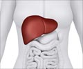
These imaging techniques helps in localising the tumors, measure the extent of spread, and to identify if the liver has been affected or not.
Echocardiography of heart: The valves of the heart are often affected by carcinoid tumors. This sign come late. Hence it is worth performing an echocardiography of heart so as to find out at the earliest, the damage that the tumor has done to the heart.


