Diagnosis of Neurocysticercosis / Cysticercosis of Brain / Pork Tapeworm
The most useful diagnostic test and the primary diagnostic criteria is neuroimaging.
Diagnosis stems from suspicions that arises from the clinical manifestations of the disease. Patients from endemic areas presenting with seizures, a normal neurological examination and spontaneous resolution after therapy with albendazole is consistent with a diagnosis of neurocysticercosis
Imaging
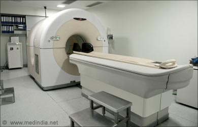
The most useful diagnostic test and the primary diagnostic criteria is neuroimaging, at first by Contrast CT (shows ring enhancement, nodular calcifications), If that is not conclusive then an MRI has to be performed. A contrast CT will pick up a calcified cyst whereas MRI is better for identifying cystic lesions and enhancement

Immunologic Assays
The second major diagnostic criteria is the detection of specific antibodies to cysticerci by using a specific immunoblot assay. However patients with single lesions and calcifications may be seronegative. But these assays are not widely available.
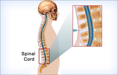
CSF examination for neurocysticercosis is indicated in patients presenting with new-onset seizures or neurological deficit whose neuroimaging does not give a definitive diagnosis.

CSF findings include the following:
- Mononuclear pleocytosis
- Normal or low glucose levels
- Elevated protein levels
- High IgG index
- Oligoclonal bands, in some cases
- Eosinophilia (5-500 cells/µL); however, this also occurs in neurosyphilis and CNS tuberculosis
- CSF ELISA for neurocysticercosis has a sensitivity of 50% and a specificity of 65%
However, these assays are not widely available.


