Diagnosis of Endometrial Cancer
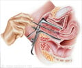
The gold standard for diagnosis of endometrial carcinoma is dilatation and curettage (D&C)
In the advanced stages, abnormalities in the uterus, mainly its size and shape may show up. Patients who report abnormal vaginal bleeding and have risk factors for endometrial cancer should have histological evaluation of the
Endometrial Sampling: A small amount of tissue is removed from the uterine wall by inserting a narrow tube into the uterus. It is usually done at the doctor’s office.
Dilatation and curettage (D & C): It involves dilating (widening) the cervix (the opening of the uterus) and inserting an instrument to scrape or suck the uterine wall and collect the tissue. D & C is also an outpatient procedure. The gold standard for a histological evaluation of the endometrium has been dilatation and curettage (D&C).
Ultrasound measurement of endometrial thickness: It has been suggested as a screening technique to obtain an image of the endometrial lining and predict the likelihood of disease based on its thickness. Measuring endometrial thickness with the use of ‘Transvaginal Ultrasonography’ has indicated that an endometrial thickness of less than 5 mm is rarely associated with carcinoma.
Sono-hysterography: This technique provides the complete characteristics of the endometrial lining and helps in differentiating between thickness, polyps and leiomyomas. Such endometrial abnormalities can easily be picked up with this technique, which might be overlooked during endometrial biopsy.
Hysteroscopy: It enables a first hand scrutiny of the endometrium, which is found to be useful in assessing abnormal uterine bleeding. This technique has an edge over Endometrial biopsy, Transvaginal


