Ventricular Septal Defect (VSD)
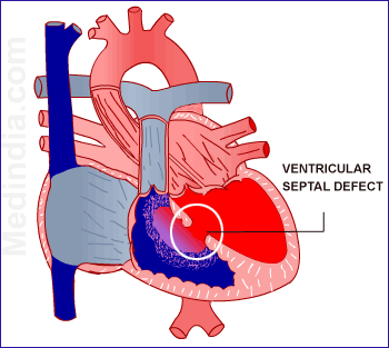
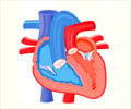
Ventricular septal defect is an abnormal hole in the septum that separates the two bottom chambers of the heart called ventricles.
Like Atrial septal defect (ASD), there are no evident reasons to say why a ventricular septal defect develops. Genetics and Chromosomal abnormalities are associated with the defect.
A ventricular septal defect allows oxygenated blood from the left ventricle to flow across to the right ventricle. VSDs can be of different sizes. Small VSDs may close on their own and usually does not cause major problems. A membranous VSD is found in the upper portion of the interventricular septum. A muscular VSD is found in the lower part of the septum. These are the kinds that can close at any time during childhood. Medium and large sized VSDs are unlikely to close spontaneously. Surgery or other interventional procedures may be required to close these defects. Inlet and Outlet VSDs are less common types and are present where the blood enters or leaves the ventricles.
Some of the infants may show poor weight gain, shortness of breath or even bluish discoloration of the lips, nails or skin. Most of the small VSDs may go unnoticed. A murmur can be heard with a stethoscope when the baby is a few weeks old.
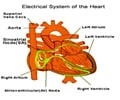
Untreated moderate to large VSDs may lead to severe complications in a child. Heart failure may result from the constant overload of the right ventricles. Arrhythmias and Pulmonary hypertension can also be an outcome of the high volume of blood flowing through the right ventricle. It is rare that these defects can go unnoticed and so complications are rather rare.






I have VSD since birth [Tetralogy of Fallot] and I'm now 29 years old, I'm aware that I'm not allowed to do any strenuous activities but is yoga allowable for me? I can't afford to go to a doctor. I'm from a 3rd world country in Asia.
HOLE IN HEART IN MY COSION HEART ,I HAVE NO MORE MONEY FOR SURGERY.MY BABY LIE ON BED AND WAITING FOR DEATH.PLEASE SUGGEST ME FOR SOME WELFARE SOSITY ,WHO HEALP MY CHILD.
dear friend, why don't u try get BPL [below poverty line]card, once this card made i belive free medicins and treatment avilable at all govt hospitals.
get it done in Sathyasai Institute of Medical sciences in Bangalore.. They will do for free..
Well and briefly explained; VSD account for upto 25% of all Cardiac Heart Failure, which simply means that 2 out of 1000 lives birth are affected. Isolated complex malformations do happened and lower left sternal edge with/or without parasternal thrill is encountered mostly during examination. Yeah/and ballabalala....
Ventricular Septal Defect Ventricular Septal Defect is usually symptomless at birth. It usually manifests a few weeks after birth. Small VSD can be asymptomatic, but larger ones can result in heart failure, pulmonary hypertension or growth restriction with recurrent respiratory infections like pneumonia. Other features may be poor weight gain, breathlessness on breast feeding and increased heart rate. If not intervened, it can develop into Eisenmenger Syndrome, which has a very bad outcome. http://heart-consult.com