Leprosy - Diagnosis
Physical examination: The doctor should look for hypo-pigmented patches or nodular lesions and also loss of sensation in the extremities.
Skin scrapings: Samples from the skin are obtained from the edge of the lesion, which is smeared on the slide and stained with Ziel-Neelsen technique. This is very helpful in demonstrating the bacilli in the skin scrapings.
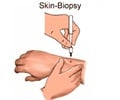
Biopsy: Biopsy of the nodular lesions, thickened nerves and lymph node puncture is useful in demonstrating the presence of the bacilli.
Lepromin skin test: It is a skin test not to diagnose leprosy but to efficiently measure the Cell Mediated Immunity (CMI) of the person. The person is injected with the antigen (lepromin A). The early reaction consists of erythema and induration, which remains for 3-5 days and is not of much significance. The late reaction commences after 1-2 weeks and reaches a peak in 4 weeks time. It consists of indurated skin nodule, which later ulcerates. It distinguishes between people who can mount a CMI response against the bacilli with those who are not capable of eliciting a response.
It is positive in case of Tuberculoid type and negative in Lepromatous Leprosy.
Antibody detection: Detection of antibody against M.leprae antigen is a very useful and specific diagnostic test.
Mouse footpad inoculation test: It is a very sensitive test for the detection of leprae bacilli in the tissue but it is unsuitable for routine diagnosis. Hence it is used only for drug resistance testing and research studies.
Advanced Molecular Testing: Polymerase chain reaction (PCR)-based methods have been useful in confirming the diagnosis in paucibacillary leprosy where few bacilli are present. It is also accurate in the detection of rifampicin/dapsone resistant strains.


