- Variations of pterygium prevalence by age, gender and geographic characteristics in China: A systematic review and meta-analysis - (http://journals.plos.org/plosone/article?id=10.1371/journal.pone.0174587)
- Prevalence and risk factors of pterygium and pinguecula: the Tehran Eye Study - (https://www.ncbi.nlm.nih.gov/pubmed/18600244)
- Test distance vision using a Snellen chart - (https://www.ncbi.nlm.nih.gov/pmc/articles/PMC2040251/)
- Ophthalmic Pterygium - (https://www.ncbi.nlm.nih.gov/pmc/articles/PMC3069871/)
- Current concepts and techniques in pterygium treatment - (https://www.ncbi.nlm.nih.gov/pubmed/17568207)
- Mitomycin eye drops as treatment for pterygium - (https://www.ncbi.nlm.nih.gov/pubmed/3211484)
- Pterygium Classification - (http://ophthaclassification.altervista.org/pterygium-classification/)
- Modified bare sclera method for the treatment of primary pterygium: a preliminary report - (https://www.ncbi.nlm.nih.gov/pubmed/15909715)
- [A brief history of diagnosis and treatment of Pterygium in China and outside China] - (https://www.ncbi.nlm.nih.gov/pubmed/24429035)
- Pathogenesis of pterygia: role of cytokines, growth factors, and matrix metalloproteinases - (https://www.ncbi.nlm.nih.gov/pubmed/15094131/)
- Management of Pterygium - (https://www.aao.org/eyenet/article/management-of-pterygium-2)
- Pterygium: Nonsurgical Treatment Using Topical Dipyridamole – A Case Report - (https://www.ncbi.nlm.nih.gov/pmc/articles/PMC3995373/)
What is Pterygium?
Pterygium or Surfer’s eye is a degenerative condition of the conjunctiva that proliferates as a fibrovascular growth to invade the cornea.
Pterygium or Surfer’s eye is referred to as a pinkish wing-shaped ocular surface lesion or a benign tissue growth onto the cornea. It is characterized by degenerative changes of the collagen and fibrovascular proliferation.
According to a given pathological theory, pterygium is typically characterized by a potentially highly proliferative tissue and reduced number of stem cells in the corneal margin. Reports indicate that it is highly prevalent in tropical areas of the world. Pterygium usually does not cause problems and does not need surgical treatment.
What are the Different Stages in Pterygium?
Pterygium does not always lead to visual impairment but if it is in an advanced form, it can certainly cause significant distortion. Pterygium has been classified in different stages that help in surgical intervention.
Stage 0: This stage is when the lesion is posterior to the limbus (border of the cornea that is contact with the white of the eye (sclera)), specifically called pinguecula. A pinguecula is a yellowish patch or bump on the conjunctiva that occurs due to a deposition of protein, fat or calcium on the tissue. In this case there is no vascularity and conjunctival and corneal ectasia are seen.
Stage 1: In this the lesion involves limbus. Minimal papillary response is seen and conjunctival and corneal tissues are flat.
Stage 2: In this stage the lesion appears just on the limbus. The vascularity is normal but a minimal elevation is observed on conjuctival and corneal tissues.
Stage 3: The pterygium covers the area between the limbus and pupillary margin. Moderate vascularity with vessel congestion is seen and the lesion is upto 1 mm.
Stage 4: In this case the lesion is central to the pupillary margin. It extends to more than 1 mm. This is a severe form of pterygium with vessel congestion and dilation. It is of dense and deep color and may involve areas of vision (visual axis). This is associated with increase in astigmatism and can even lead to limitation of eye movement.
What are the Types of Pterygium?
A pterygium can either be progressive or atrophic (degenerated)
The differences between the two are -
| Progessive pterygium | Atrophic pterygium | |
| Appearance | Thick and fleshy | Thin and membranous |
| Blood vessels | Very prominent | Very few blood vesssels giving a pale appearance |
| Cap in front of the head | Present | Absent |
| Progression | Continues to advance further into the cornea | Static after an initial period of growth |
Morphology of Pterygium
Stocker’s line - A line of iron deposition adjacent to the head of the pterygium. It appears in case of chronic pterygium.
What is the Appearance of a Pterygium?
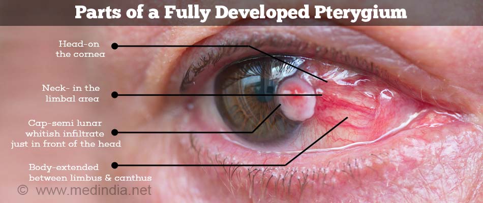
A pterygium appears as a wing shaped growth on the visible part of the sclera in the horizontal meridian, which is seen to be invading into the adjacent cornea. The color depends on the degree of prominence of blood vessels - it may be red, thick and fleshy in a progressive pterygium, and pale, thin and membranous in an atrophic pterygium. A line of iron deposition known as Stocker’s line is often seen in front of the head of long standing pterygia.
A pterygium conisists of the following parts -
- Head: Part of the pterygium in the cornea
- Neck: the part that overlies the junction between the cornea and sclera
- Body: the part that lies in the conjunctiva overlying the sclera
- Cap: opaque infiltration seen in front of the head of progressive pterygia

What are the Causes of Pterygium?
The exact cause of pterygium is not clear but most of the experts believe that long-term exposure to ultraviolet light (UV-B rays specifically) plays an important role. Exposure to wind, chemicals, pollution and eye irritation from dry, dusty conditions may also lead to development of pterygium. The risk of pterygium is higher in tropical areas or ozone layer-depleted regions of the world, which have reduced ultraviolet light filtering. It is believed that there is a dysfunction in the stem cells situated near the corneo-scleral junction which results in the formation of a pterygium. Hence most of the treatments address this issue by attempting to replace the abnormal stem cells in this region with normal stem cells.

What are the Symptoms of Pterygium?
Usually pterygium is asymptomatic apart from its appearance.
Sometimes a pterygium can give rise to the following symptoms -
- Dryness, grittiness or foreign body sensation - Due to rapid tear evaporation because of an uneven ocular surface.
- Diminished vision - Either due to the growth altering the shape of the cornea producing astigmatism, or due to obstruction of the visual axis.
- Redness and pain - If the pterygium is inflamed.
- Diplopia - This is very rare, and occurs in very large recurrent ptergia due to restriction of ocular movments.
How do you Diagnose and Evaluate Pterygium?
Special tests are not necessary for diagnosing a pterygium. An ophthalmologist generally performs a physical examination using a slit lamp to diagnose the condition. A slit lamp allows the doctor to observe the eye using magnification and bright lighting.

Visual acuity is checked and refraction is performed to determine the presence and type of any refractive error. The degree of invasion of the cornea, and whether the visual axis is threatened is determined, The presence of any inflammation is noted.
Photo documentation may be done. It involves taking pictures to track the growth rate of the pterygium. A clinical record is maintained so that, when the patient visits the doctor the next time, it can be compared with previous photographs to see if the pterygium has grown.
How do you Treat Pterygium?
An asymptomatic pterygium need not be treated. A symptomatic pterygium may be managed in one of the following ways –
Medical managemnt
- Ocular lubricants in the form of eye drops for dry eye symptoms
- Spectacles for mild cases of astigmatism
- Steroid eye drops for inflamed pterygia
Surgical management
Surgical removal is indicated in the following conditions -
- For cosmesis
- Progressive pterygium threatening to encroach the visual axis
- Large Pterygium causing severe astigmatism
- Repeated episodes of inflammation
- Double vision because of the pterygium
Since surgical excision in isolation is known to be followed by recurrence of the pterygium in almost all cases, one of the following additional measures needs to be taken to prevent recurrence. These are –
- Conjunctival autograft: removing a small part of the conjunctiva either from the same or opposite eye and placing it in the area of defect caused by the removal of the pterygium. A conjunctival graft provides normal stem cells to promote normal conjunctival growth.
- Amniotic membrane grafting: The defect is covered by amniotic membrane obtained from a placenta. The amniotic membrane provides normal stem cells to promote normal conjunctival growth.
- Mitomycin C: This is an agent that inhibits the growth at the conjunctival level and thus helps in prevention of recurrences. Either it is placed on the bed of the sclera after removal of the pterygium, or it is administered in the form of eye drops after the surgery.
If there is extensive scarring of the cornea in the visual axis, a partial thickness corneal grafting may have to be combined along with the other surgical procedures.
How do you Prevent Pterygium?
In order to prevent a surfer’s eye formation, or reducing the risk of a pterygium recurrence, the following measures can be adopted.
- Use sunglasses to block ultra-violet rays.
- Wear a hat whenever out in sun.
- Avoid exposure to environmental irritants such as smoke, dust, wind and chemical pollutants as much as possible.
- Use appropriate eye safety equipment in the work place.
- Patient counseling should be done before and after surgery.


