Risk Factors
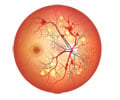
- Lattice degeneration is a type of thinning of the peripheral retina which predisposes to retinal detachment. It is seen in 10% of the population and fortunately only 1% progress to retinal detachment. It is called lattice because the area of thinning has white cris-cross lines over it resembling a lattice. The vitreous gel is unsually firmly attached to this area of thinning, hence its separation from the retina results in a tear leading to retinal detachment. Lattice degeneration is more commonly seen in high myopic people (short sighted people)
Other degenerations like snail track, meridional folds also rarely predispose to retinal detachment.
- High myopic individuals have a higher incidence of retinal detachment. The prevalence appears to be related to the severity of myopia.
- Patients with chronic uveitis (long standing inflammation of the eye) are prone to retinal detachment.
- Patients with advanced stages of diabetic eye disease are at a high risk of retinal detachment.
- Retinopathy of prematurity affecting children on high concentrations of oxygen following premature birth.
- Chronic infections like tuberculosis and some autoimmune diseases can also lead to a retinal detachment.


