- Central Retinal Vein Occlusion - (https://www.ncbi.nlm.nih.gov/books/NBK525985/)
- Retina Health Series - (https://www.asrs.org/patients/retinal-diseases/22/central-retinal-vein-occlusion)
- Branch Retinal Vein Occlusion - (https://www.asrs.org/patients/retinal-diseases/24/branch-retinal-vein-occlusion)
About
Retinal Vein Occlusion (RVO) is a serious eye condition that occurs when one of the veins responsible for draining blood from the retina becomes partially or completely blocked. The retina, a delicate layer of tissue at the back of the eye, is crucial for translating light into the images we see. When a retinal vein is blocked, it disrupts the blood flow, leading to complications such as increased intraocular pressure and retinal swelling. These complications can result in significant vision loss if not promptly treated(1✔ ✔Trusted Source
Central Retinal Vein Occlusion
Go to source).
Retinal Vein Occlusion is the second most common retinal vascular disorder, following diabetic retinopathy. It affects over 16 million people worldwide. Central Retinal Vein Occlusion affects between 1 and 4 per 1,000 people, while Branch Retinal Vein Occlusion is more common, affecting between 6 and 12 per 1,000 people.
Did You Know?
RVO is the second most common retinal vascular disorder, affecting over 16 million people globally. #visioncare #medindia
Types of Retinal Vein Occlusion
RVO is categorized into two main types:
- Central Retinal Vein Occlusion (CRVO): This type involves a blockage in the main retinal vein, leading to more severe consequences(2✔ ✔Trusted Source
Retina Health Series
Go to source). - Branch Retinal Vein Occlusion (BRVO): This occurs when one of the smaller branch veins is blocked. BRVO is more common than CRVO but typically less severe(3✔ ✔Trusted Source
Branch Retinal Vein Occlusion
Go to source).
Symptoms and Signs of Retinal Vein Occlusion (CRVO and BRVO)
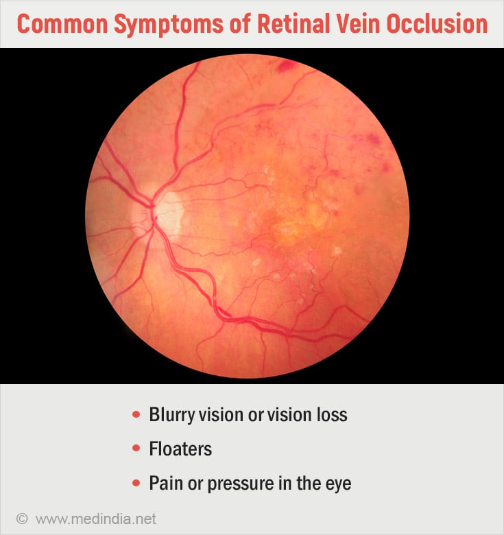
Retinal Vein Occlusion, whether central (CRVO) or branch (BRVO), often presents with subtle yet significant symptoms. Both CRVO and BRVO share some symptoms and signs, such as vision changes, floaters, and retinal hemorrhages. The extent and severity depend on the location and size of the blocked vein.

Central Retinal Vein Occlusion (CRVO)
CRVO occurs when the central retinal vein, located at the optic nerve, becomes blocked.
Symptoms:
Sudden, painless vision loss: This can be partial or complete, and it often affects only one eye.
Blurry or distorted vision: Objects may appear blurred or wavy.
Dark spots or floaters: Patients may see dark spots or streaks in their vision.
Signs:
Retinal hemorrhages: Blood may appear in multiple areas of the retina, seen as blotches.
Optic disc swelling: The optic nerve may appear swollen on examination.
Cotton wool spots: These are fluffy white patches on the retina, indicative of nerve fiber damage.
Macular edema: Swelling in the central part of the retina, leading to vision distortion.
Branch Retinal Vein Occlusion (BRVO)
BRVO occurs when one of the smaller branches of the retinal vein is blocked.
Symptoms:
Peripheral vision loss: This is more common than central vision loss and may affect part of the visual field.
Blurry vision: Similar to CRVO but typically less severe and localized to the area of the blockage.
Floaters: Small spots or strings that float through the vision.
Signs:
Localized retinal hemorrhages: Bleeding is confined to the area supplied by the blocked vein.
Macular edema: Swelling in the macular can cause central vision distortion.
Venous dilation and tortuosity: The affected vein may appear enlarged and twisted.
Causes of Retinal Vein Occlusion
The underlying cause of RVO is a disruption in the normal blood flow through the retinal vein. This disruption can result from:
Blood Clot Formation: A clot can obstruct the vein, leading to RVO.
Slowed Blood Flow: Reduced blood flow can increase the risk of blockage.
Compression of the Retinal Vein: The retinal artery, which supplies oxygen-rich blood to the retina, can become stiff due to aging or plaque buildup. This stiffness can compress the adjacent retinal vein, causing damage to the vein's inner lining and increasing the likelihood of a clot forming.
Risk Factors for Retinal Vein Occlusion
Several factors can increase the risk of developing RVO:
Age: The risk of RVO increases with age, particularly in individuals over 40. Most cases occur in people in their 50s or 60s, though it can affect younger individuals as well.
Medical Conditions: Certain health conditions, such as atherosclerosis, diabetes, glaucoma, and high blood pressure, can elevate the risk of RVO.
Previous RVO: A history of RVO in one eye increases the likelihood of it occurring in the other eye.
Complications of Retinal Vein Occlusion
RVO can lead to several serious complications, including:
- Cystoid Macular Edema: Swelling in the macula, the central part of the retina, can cause blurry vision or vision loss.
- Neovascularization: Abnormal blood vessels may form in the eye, particularly on the iris (rubeosis iridis) or retina, leading to further complications.
- Vitreous Hemorrhage: Bleeding into the vitreous humor, the gel-like substance filling the eye, can occur due to the formation of abnormal blood vessels.
- Neovascular Glaucoma: Abnormal blood vessel growth can increase intraocular pressure, leading to glaucoma.
- Retinal Detachment: The formation of abnormal blood vessels in the retina can cause it to detach from the supporting tissues.
Moreover, individuals with RVO have a higher risk of developing cardiovascular diseases, including stroke, due to shared risk factors such as high blood pressure and atherosclerosis.
Diagnosis of Retinal Vein Occlusion
Eye care specialists diagnose RVO through a combination of eye exams and advanced retinal imaging techniques. Coordination with a primary care physician (PCP) is essential to identify the underlying causes of blood flow problems.
During the eye exam, the specialist will dilate the patient’s pupils to examine the back of the eye using a microscope and ophthalmoscope. This thorough examination helps identify signs of macular edema, abnormal blood vessel formation, and the extent of retinal ischemia.
Diagnostic Tests for Retinal Vein Occlusion
Several tests may be conducted to diagnose RVO and assess the severity of the condition:
Fundus Photography: This imaging technique helps visualize abnormal blood vessels and the extent of bleeding within the eye. Fundus Imaging Findings are:
- "Blood and Thunder" Appearance: Commonly seen in CRVO, the retina may show extensive hemorrhages giving it a dramatic appearance. BRVO, while also showing hemorrhages, usually has a more localized presentation.
- Cotton Wool Spots and Optic Disc Swelling: These findings are typical of both conditions, indicating damage to the retinal nerve fibers.
- Non-ischemic CRVO usually presents with mild dilation and tortuosity of the veins, while BRVO findings are localized based on the affected branch.
- Ischemic CRVO is marked by more severe retinal edema and extensive hemorrhages, which are not typically as pronounced in BRVO.
Optical Coherence Tomography (OCT): OCT provides high-resolution images of the retina, allowing the specialist to detect macular edema and measure retinal thickness accurately. This information is crucial for guiding treatment decisions.
Fluorescein Angiography: In this test, a dye is injected into a vein in the arm, which then travels to the retinal blood vessels. The dye highlights any blockages or areas of reduced blood flow in the retina, providing detailed images that aid in diagnosis and treatment planning.
Management and Treatment of Retinal Vein Occlusion
Management of RVO requires a multidisciplinary approach, with eye care specialists and primary care physicians working together. Blood tests may be necessary to assess cholesterol levels, blood sugar, and other factors that could contribute to the condition.
Treatment Approaches for Retinal Vein Occlusion
Currently, there is no cure for RVO, but treatment focuses on managing complications and preserving vision. Treatment options include:
Anti-VEGF Injections: These injections are the first-line treatment for macular edema associated with RVO. VEGF (vascular endothelial growth factor) promotes the growth of abnormal blood vessels that can leak and cause swelling. Anti-VEGF injections help reduce VEGF production, thereby reducing swelling and preventing further vision loss. Patients may require regular injections over one to two years.
Common medications used in these injections include:
- Aflibercept
- Bevacizumab
- Ranibizumab
Steroid Injections: Intraocular Steroid injections like, either a liquid steroid called triamcinolone or a small steroid pellet called dexamethasone implant can also reduce retinal swelling. However, they are typically used as a second-line treatment due to potential side effects, such as elevated intraocular pressure and cataract formation.
Panretinal Photocoagulation (PRP): This laser surgery creates small burns in areas of the retina that lack blood flow. By reducing the production of VEGF, PRP helps prevent neovascularization and stabilizes intraocular pressure.
Vitrectomy Surgery: Vitrectomy is a surgical procedure recommended for severe cases of RVO, particularly when there is significant bleeding (vitreous hemorrhage) or retinal detachment. The surgery involves removing the vitreous humor and repairing retinal damage.
Many individuals with RVO have underlying conditions such as high blood pressure, diabetes, or high cholesterol. Managing these conditions is crucial to reducing the risk of further complications.
Preventing Retinal Vein Occlusion
While it may not be possible to prevent RVO entirely, individuals can take steps to reduce their risk by addressing the underlying causes of blood flow problems. Preventive measures include:
- Adopting a diet that supports heart and blood vessel health can lower the risk of RVO.
- Incorporating physical activity into daily routines helps maintain cardiovascular health and manage weight.
- Maintaining a healthy weight is crucial for overall health and reducing the risk of RVO.
- Smoking and other forms of tobacco use increase the risk of blood vessel damage and should be avoided.
What to Expect with Retinal Vein Occlusion
The prognosis for individuals with RVO varies depending on the location and severity of the blockage and the presence of complications. Some people may experience permanent vision damage, while others may see gradual improvement over time.
Your eye care specialist is the best resource for understanding your specific prognosis and what to expect as you manage the condition.
In some cases, vision rehabilitation may be recommended. This form of rehabilitation helps individuals adapt to reduced vision through techniques such as using magnifying devices or assistive technology. Social workers and other professionals can also provide support to help individuals cope with lifestyle changes resulting from vision loss.
Living with RVO can be challenging, but with proper management and support, many individuals can maintain a good quality of life. Regular follow-ups with an eye care specialist, adherence to treatment plans, and addressing underlying health conditions are essential for managing the condition and preserving vision.


