- Chest X-Ray - (http://www.nlm.nih.gov/medlineplus/ency/article/003804.htm)
- Dick E. Chest radiographs made easy. Western Journal of Medicine 2002;176(1):56-57.
- About Chest X-Ray - (http://www.nhlbi.nih.gov/health/health-topics/topics/cxray)
What is Chest Radiography?
Chest radiography is a commonly used imaging diagnostic tool. It involves the use of chest radiographs, more commonly referred to as chest x rays. This is a widely used method of diagnosis for a wide range of conditions as it provides health care experts with images of the heart, lungs, large arteries, chest bones and spine. A chest radiograph can also reveal the presence of fluid in or around the lungs.
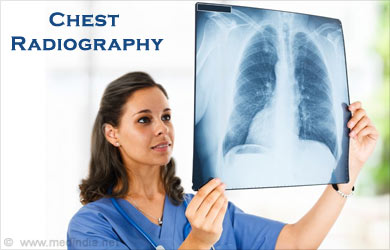
Chest radiography is commonly used not only in the diagnosis of various health conditions, but also used to monitor the progress of treatment. In such scenarios, a series of chest X-rays may be produced over a specific period of time to help track progression of a disease and response to treatment.
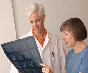
Chest X-ray films may not appear to reveal much when examined by a lay person, but this is because the individual is not trained to interpret the results. Technicians at the laboratory and doctors will be able to interpret chest X-ray results and give a better insight into what the test reveals.
When Do You Need a Chest X-Ray?
As is the case with any diagnostic test, you do not need to and should not get a test done unless your doctor has advised you to do so. Your health care provider will determine when an X-ray is required and will advise you accordingly. Doctors generally ask patients to first undergo chest radiography if they suspect any heart or lung disease, or if there is an injury in the region. There are certain circumstances under which you may be asked to do a chest x ray and these are:
- If you have suffered from a persistent cough that does not respond to conventional treatment.
- If you have suffered an injury to the chest.
- If you have been experiencing chest pain and the cause is unknown.
- If you have been experiencing difficulty breathing.
- If traces of blood have been observed in the sputum or phlegm.
How Does a Chest X-Ray Help?
Chest radiography will help your health care provider arrive at a more accurate diagnosis, which is obviously the key to a successful treatment of any health condition. In other cases, it will help to rule out certain possible health conditions.
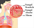
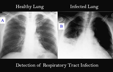
- Lung Health: A chest radiograph will help in the detection of cancer, a respiratory tract infection, the accumulation of fluid in and around the lungs or pneumothorax, which is an abnormal accumulation of air in the area around a lung. It can also help reveal the presence of chronic lung diseases like cystic fibrosis or emphysema and other complications.
- Heart Disease: A chest radiograph can be used to find changes in the shape and size of the heart that may be indicative of heart failure, pericardial effusion or heart valve problems. In certain cases respiratory problems may also result from heart problems such as the accumulation of fluid in the lungs resultant from congestive heart failure. Chest x rays can also reveal heart damage that occurs as a result of calcium deposits.
- Blood Vessel Problems: X-ray images give doctors an insight into the health of the larger blood vessels around the heart such as the aorta and pulmonary arteries and veins. This can help to identify certain blood vessel problems, aortic aneurysms and congenital heart disease.
- Physical Injuries: Any disease or injury that causes skeletal changes can be picked up through chest radiography. Rib or spine fractures are commonly identified from chest X-rays.
- Postoperative and Post Treatment Use: Chest X-rays are routinely used after the medical devices like catheters, pacemakers and defibrillators are installed just to make sure that the procedures were successful. Similarly X-rays are used after surgeries to treat heart, lung or esophageal disorders.
How to Prepare for a Chest Radiograph and What You Can Expect?
You can get a chest X-ray done at any clinic or diagnostic center, in most hospitals and even in some doctors’ clinics. When you go in for a chest X-ray you will first need to remove all clothing and jewelry from the waist up. This is necessary as both fabrics and jewelry can obscure the results of X-ray imaging.
- You will be asked to stand in front of the machine and hold your breath while the X-ray is being taken.
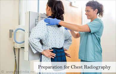
- More often than not an X-ray image is taken from 2 angles.
- In this case, you will first be asked to stand facing the machine and then sideways.
- You will not experience any discomfort while undergoing a chest X-ray.
Make it a point to inform your health care provider if you are pregnant. Your health care provider will advise you accordingly, but in most cases chest X-rays are not conducted during the first two trimesters of a pregnancy.
Understanding the Risks
It is understandable for one to be concerned about the risk of exposure to radiation from X-rays, but the risk is in fact extremely low. Radiography machines are monitored regularly and have to meet stringent safety requirements. They utilize the minimum amount of radiation that is required to obtain your radiograph image. Moreover, the minimal risks presented from this form of radiation far outweigh the benefits. The risk is obviously higher if you undergo radiography regularly. Under normal circumstances, X-rays are only required occasionally and therefore present no real danger.
Pregnant women and children are not advised to undergo X-rays unless absolutely necessary as the risks are higher for them.

