- Urinary Tract Imaging - (http://www.niddk.nih.gov/health-information/health-topics/diagnostic-tests/imaging-urinary-tract/pages/imaging-urinary-tract.aspx)
- Your Kidneys & How They Work - (http://www.niddk.nih.gov/health-information/health-topics/anatomy/kidneys-how-they-work/pages/anatomy.aspx)
- Nuclear Medicine - (http://www.nibib.nih.gov/science-education/science-topics/nuclear-medicine)
- Short Term Prognostic Utility of Tc-99m DMSA Renal Imaging in Sepsis Induced Acute Renal Failure; Provisional Data - (http://dx.doi.org/10.4236/ijcm.2013.412094)
What is Renal Scan / Isotope Renogram / Renal Scintigraphy?
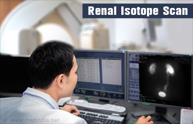
Renal Isotope Scan or an Isotope Renogram is a diagnostic procedure done by nuclear medicine specialists using a radioactive tracer to study the structure and function of the kidneys.
It takes anything from 1 to 3 hours to perform and is done by a specialist radiologist in a diagnostic center or a hospital. You or your doctor will need to book an appointment to have this test done.

How do Kidneys Remove Wastes from the Body?
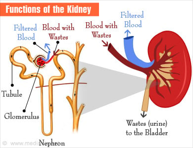
The kidneys remove waste material like urea, uric acid and creatinine from the blood, and maintain water and electrolyte levels in the body. Each of the two kidneys has a million units called nephrons. Each nephron consists of a filtering unit called glomerulus, and proximal and distal tubules. The distal tubule opens up into collecting ducts, which join to form a single duct that takes the urine out of the kidneys into the ureter.
Blood from the body is brought to the glomeruli by branches of the renal artery, where fluid and waste material are filtered into the proximal tubule.
The filtered material passes through the proximal and distal tubules where selective reabsorption and secretion of certain substances, water and electrolytes take place. It then passes into collecting ducts, and finally out of the kidney into the ureters and urinary bladder. Once the bladder gets full, the patient gets an urge for passing urine, which is excreted through the urethra.

How does a Renogram / Renal Scan / Renal Scintigraphy Work?
During a renal scan, a small amount of radioactive substance called a radiotracer is injected into a vein. Technetium 99m is a commonly used radioactive tracer to assess kidney function. As it passes through the body, it emits gamma rays, which can be detected using a gamma camera.
The radioactive tracer has to be guided to the kidneys so that kidney function can be assessed. For this reason, it is attached to a carrier molecule like DMSA (2, 3-dimercaptosuccinicacid), diethylenetriamine pentaacetic acid (DTPA), mercaptoacetyltriglycine (MAG3), or iodine-131 or iodine-123 orthoiodohippurate (OlH), which takes it to the kidneys. The preferred carrier molecule is determined by the suspected underlying kidney pathology.
This guiding molecule differs from organ to organ. For example, ligand methylene-diphosphonate (MDP) is preferentially taken up by bone and attaching it to technetium-99m, the radioactivity can be transported to bones for a scan to detect tumor or fractures.
The radiotracer emits gamma rays from the kidneys, which are detected with a special scanner called gamma camera. The gamma camera converts the signals into electric signals that are passed on to a computer to create images of the kidneys, which are interpreted by a nuclear medicine specialist.
Diuretic Renogram - Some patients may also receive diuretics like furosemide before the procedure. Diuretics are used in cases of hydronephrosis or dilatation of the upper urinary tract. They help determine if an obstruction is responsible for the pathology.
ACE inhibitor scintigraphy- Patients with uncontrolled blood pressure may be advised a procedure called ACE inhibitor scintigraphy. This test helps determine if the cause of the Know About High Blood Pressure is at the level of the kidneys at the renal artery, a condition referred to as renovascular hypertension.
Patients with renal artery stenosis have reduced blood supply to the kidney. This stimulates a hormonal system called the renin-angiotensin-aldosterone axis, resulting in high blood pressure. Though ACE inhibitors (angiotensin converting enzyme inhibitors) like lisinopril and ramipril are commonly used antihypertensive drugs, these patients worsen when administered these drugs. The ACE inhibitor renal scan is taken before and after administration of an ACE inhibitor, usually captopril.
What are the Common Uses of a Renal Scan / Renal Scintigraphy?
There are several radiological tests used to detect kidney problems like CT scan, MRI and ultrasound. Depending on the suspected problem, your doctor may require an isotope renal scan. An isotope renal scan detects problems with glomerular filtration and with renal tubular function or both. It can be used for the following condition:
- To assess function of a kidney if it has suffered an injury or an insult or before removing it for transplantation in a kidney donor.
- To assess perfusion and function of a transplanted kidney.
- To check scars on the kidney due to infections.
- To check if kidneys are responsible for high blood pressure that does not respond to routine medications.
- To determine the cause of urinary tract dilatation like pelvi-ureteric junction or PUJ obstruction.
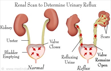
- To determine if there is reflux in the urinary tract.

How Should the Patient Prepare for the Procedure?
You may have to undergo other tests like ultrasound and urine test before undergoing renal scintigraphy. When your doctor advises you to undergo a renal scintigraphy:
- Allergies - Inform your doctor if you suffer from any allergies. Some people may be allergic to the radiotracer injected during the scan. However, the reaction is usually minor even if it is present.
- Pregnancy - If you are a woman, inform the doctor if you are pregnant.
- Illness or Medication - Inform your doctor if you have recently suffered any illness or if you are taking any medications
- Jewellery - Do not wear jewellery during the test
- Stop ACE inhibitor tablets - If you are undergoing an ACE inhibitor renal scintigraphy and you are taking an ACE inhibitor for your blood pressure, it will have to be stopped a few days before the procedure.
What Happens during a Renal Scan?
The radionuclide scan does not require prior admission to a hospital. However, the test could take a few hours and you will need to be prepared for it. Be sure you are well hydrated before the procedure.
Intravenous line - When you arrive for the test, a nurse will place an intravenous line in a vein of your arm or hand, into which the radionuclide will be injected.
Fluids may be injected into your vein following the injection.
You will be asked to wait for a variable time of between half an hour to 3 hours so that the radionuclide reaches the kidneys, before the scanning begins.
You may also be administered furosemide or captopril depending on the type of scan.

Once the necessary time has lapsed, you will be asked to lie down on the scan table with the big special gamma camera.
You will have to remain still while the pictures are being taken. You may be asked to shift your position at regular intervals. You may also be asked to go to the restroom to empty your bladder in between, and also after the procedure.
What is the Difference between Dynamic and Static Scintigraphy?
In dynamic renal scintigraphy, a series of images are obtained almost continuously as the tracer passes through the urinary tract. Thus, it can show the function of each component of the kidney separately.
In static scintigraphy, images are taken within a particular time frame, for example, 2 to 3 hours following the injection. It is mainly useful to evaluate structural problems of the kidney. Any damaged area due to conditions like pyelonephritis or renal scars due to trauma will appear as areas of abnormal or irregular uptake of the radiotracer. This type of scan is sometimes called a DMSA scan.
What Happens after the Procedure?
Once the scan is complete, you may be asked to sit in the office for some time before you go home.
You will be asked to drink plenty of water so that you feel the urge to pass urine frequently, and the radiotracer can be washed out from the body in the urine.
If you were administered an ACE inhibitor, your doctor would confirm that your blood pressure is normal before you leave the scanning center.

How does the Doctor Interpret the Scan?
During the scan, the scanner converts the information into an electric signal and passes it on to a computer.
The increased accumulation of the radionuclide in certain areas of the kidney appears as hot spots, while cold spots indicate areas of low activity.
If adequate amounts of radiotracer do not appear in the kidney tubules, it means there is a problem with the glomerular filtration or no blood supply to the kidney. The experienced doctor studies the distribution of the radionuclide on the computer and interprets the scan.
What are the Complications and Risks?
A renal scan is usually without complications. However, it should be avoided in pregnant women and should be done with caution in allergic individuals.
When ACE inhibitors are administered, there could be a fall in blood pressure. Therefore, the blood pressure has to be regularly monitored during and after the procedure.


