What is Ureteroscopy?
Ureteroscopy is an endoscopic procedure that leaves no scars.
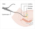
Ureteroscopy is an endoscopic procedure that is carried out to view the inside of the ureter. Ureteroscope is the instrument used for the procedure. This is a very fine instrument that is made of either a semi-rigid long steel shaft or it maybe flexible.
Ureteroscopy was developed in the 1970s and came into wide use during the 1980s. Previously a type of treatment called "blind basketing" was often used. A small basket-like device was passed — blindly, with no viewing instrument — through the urethra and bladder and into the ureter to pull out the stone. This type of "blind" treatment risked injury to the ureter and was less effective than other methods used today.
The procedure is usually done to eliminate a stone either in the ureter or in the kidney by a surgeon called Urologist.
The flexible instrument is usually used for kidney stones that are located in the lower portion of the kidney. The rigid instrument is used to remove stones or other obstructing pathologies from the ureter.
Sometimes the instrument is also used to find the cause of bleeding that is difficult to diagnose using the conventional tests.
Once the stone is identified, it can be removed totally or if it is too large to be removed , it can be broken down into smaller fragments using appropriate devices like a laser or a fine- fragmenting instrument. These fragments can be easily picked by a small, grasping instrument and be removed.
After the stone has been removed, a stent (a synthetic tube which is left in between the kidney and the urinary bladder) is left in the ureter for a few days to allow healing.
It is possible for the stent to be provided with a nylon suture attached at the distal end. This is called a dangler and the fine nylon may dangle out of your penis. This facilitates its removal without the need for any additional procedure like a cystoscopy.
The stent can be left inside the system for a few days to 2 months depending on the complexity of the procedure. It is very important for this stent to be removed because if it is left in the urinary system for a longer time, stone can form on it.
A urinary catheter is kept after the procedure to drain the urine. The majority of ureteroscopic procedures can be performed as day surgery and most individuals can return to work within a few days following the procedure.
Instruments and accessories for ureteroscopy have improved over the years. Ureteroscopes have become thinner in diameter with better fibre-optics that provides excellent view of the inside of the organ. The ureter is a very delicate thin tubular structure that connects the kidneys to the bladder and requires gentle handling. It is about 25 cms or twelve inches in length.
Before the advent of ureteroscopes, the surgeons usually had to do an open operation that left the patient with a five to six inches scar and a long recovery time of a week or 10 days. With ureteroscopy the patient is able to return home either on the same day or in a day or two. The procedure is usually done under general or spinal anesthesia.
Ureteroscopy - Anatomy & Function
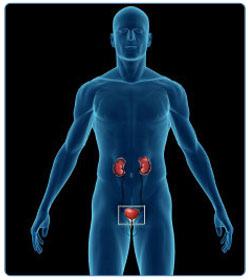
The organs, tubes, muscles, and nerves that work together to make, store, and carry urine make up the urinary system. The urinary system consists of two kidneys, two ureters, the bladder, and the urethra.
"Bones can break, muscles can atrophy, glands can loaf, even the brain can go to sleep without immediate danger to survival. But should the kidneys fail...neither bone, muscle, gland nor brain could carry on."Dr. Homer Smith in his book, "Fish to Philosopher."
Kidneys perform vital functions and many other organs in our body depend on the kidneys in order to function. A person normally has two kidneys, one on either side of the back under the lower ribs. They are shaped like kidney beans hence called “kidneys”. Each kidney is about 4 to 5 inches long and 2 to 3 inches wide. Each kidney is made up of small, complex units called nephrons. The two kidneys contain about two million nephrons. The nephrons work continuously to filter out waste products from the blood stream, all of which come from the food that one eats and the fluid that one drinks.
Kidneys are the master chemists of the body and regulate the amount of water that should stay in the body. The chemicals and wastes that accumulate have to be discarded and kidney tends to do this very precisely. Normally 200 liters of water are filtered through the kidney daily and only about 2 liters are passed as urine.
From the kidneys, urine travels down two thin tubes called ureters, leading away from each kidney to the urinary bladder. The ureters are about 24 – 30 cm long and the thickness is about 3 mm. The muscles in the walls of the ureter constantly tighten and relax to empty the urine from the kidneys. Small amounts of urine are emptied into the bladder from the ureters about every 10 to 15 seconds.
Ureter is divided into upper, middle and the lower third. It has a long course in the body by the side of the spinal cord. It has three narrow areas where the stones tend to get stuck. These narrow areas include –
- The junction between the pelvis of the kidney and the ureter.
- The middle third where it crosses over large vessels of the pelvis.
- The lower third – where the ureter meets the urinary bladder.
The urinary bladder is responsible for storing the urine produced and for letting it out at regular intervals. It sits in your pelvis and it stores urine until you are ready to empty it. It swells into a round shape when it is full and gets smaller when empty. If the urinary system is healthy, the bladder can hold up to 450 – 500 ml of urine comfortably for 2 to 5 hours.
The nerves in the bladder tell you when it is time to urinate (empty your bladder). As the bladder first fills with urine, you may notice a feeling that you need to urinate. As the bladder fills with more and more urine over a period of time, sensation to urinate becomes stronger. At that point, nerves from the bladder send a message to the brain that the bladder is full, and your urge to empty your bladder intensifies. After the brain has received the signal, it sends back an message to the urinary bladder whether it can let out the urine or not. For example, if you feel like passing urine when you are in a important meeting, you will be able to control your desire to urinate or the urge to pass urine to some degree until it is convenient to pass urine. This is because the brain has given a signal to wait for some time till it is convenient to pass urine.
When it is convenient, the brain signals the muscles of the urinary bladder to tighten which would help to squeeze the urine into the urethra through which the urine is taken out from the body.
Ureteroscopy - Ureteral Stone
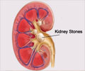
Urinary stones are gravel- like collections of various substances within the urinary tract. Stones nearly always form in the kidney, where they may remain without symptoms and do not require treatment. A ureteral stone is usually a stone from the kidney that has moved down into the ureter.
The stone begins as a tiny grain of undissolved material located where urine collects in the kidney. Over time, more undissolved material is deposited and the stone becomes larger. The material deposited is usually a mineral called calcium oxalate. Other less common materials that may also form a stone include calcium phosphate, uric acid, infected or struvite stones and cystine stones.
The stones may cause obstruction or break loose and try to pass with the normal flow of urine via the urinary tract to the outside. Most stones enter the ureter when they are still small enough to move down into the bladder. From there, they pass out of the body with urination. However if the stone is larger than the diameter of the ureter they get stuck in the narrow passage and cause excruciating pain ,called a colic, and obstruction. If this happens, treatment is required and, for some stones, ureteroscopy is the best form of treatment.
Ureteroscopy - Symptoms
Ureteric colic, due to the stone causes excruciating and unbearable pain and may last several minutes or hours.
The pain of a kidney stone has been described as the most severe pain that a person may experience. It has been described as being worse than child birth.

The pain often begins suddenly as the stone moves in the urinary tract causing irritation and blockage. Typically, the person feels a sharp and twisting type of pain in the area of the kidney or in the lower abdomen, which may spread to the groin. The pain may be steady or come in waves and may be associated with nausea, vomiting, and blood in the urine. In a man, pain may move down to the side of the inner side of scrotum whereas in women it may radiate to the inner side of the vulva. If the stone is in the lower ureter or in the bladder the pain maybe felt on the tip of the penis.
If the stone is in the lower end of the ureter, at the point where the ureter enters the opening of urinary bladder, one may feel the need to urinate more often or feel a burning sensation during urination.
If fever or chills accompany any of these symptoms, then there may be an infection. One should contact an urologist immediately as this is an absolute emergency and even more so if you have associated diabetes.
Occasionally, stones do not produce any symptoms. But while they may be "silent," they can be growing, even causing damage to the function of the kidney. If the stone is too large to pass easily, pain continues as the thin muscular wall of the ureter try to squeeze the stone along into the bladder.
It has been found out that even small stones can cause troublesome symptoms. Usually stones smaller than 4 mm have a tendency to pass out spontaneously however stones as small as 2 mm have caused symptoms.
Ureteroscopy - Investigations
A Plain CT scan is the best single investigation to confirm the presence of a ureteric stone.

The usual initial investigations to confirm the presence of a ureteric stone include –
- Routine urine test to look for presence of RBCs or red blood cells in the urine. This is due to abrasion caused by the spikes of some stones like calcium oxalate.
- An ultrasound of the kidney, ureter and bladder – to look for stones and swelling of the kidney or hydronephrosis ( hydro- water, neprhrosis – kidney).
If the ultrasound indicates presence of any hydronephrosis of the kidney but if the stone is not seen, than a plain X-ray is suggested. A stone that has calcium is usually seen on X-ray, however some stones like the ones with uric acid may not be picked up on a X-ray. Depending on the practice of the hospital and the urologist in such an event an Intra-Venous Pyelography (IVP) or a plain CT scan maybe done.
Other tests that maybe done include-
- Complete blood count and Hemoglobin
- Biochemical parameters – Electrolytes, Urea and creatinine
- Blood sugar or glucose level
- If surgery is planned a chest X- ray and an ECG maybe required.
Ureteroscopy - Types of Treatment
Ureteric stone can be treated using an ureteroscope or by ESWL (Extra Corporeal Shock Wave Lithotripsy).
Treating ureteric stones depend largely on the size, position and hardness of the stones. Luckily, the majority of small stones of about 2 to 4 mm in diameter that do not cause infection, blockage or symptoms will pass if you simply drink plenty of fluids every day. Recent studies have suggested that the majority of stones (95 percent) that are capable of spontaneous passage will pass within six weeks.
Consuming about 2 .5 to 3 .5 litre of water increases urine production, which eventually washes kidney or other stones out of the system. Once they have passed, no other treatment is necessary except continuing a high fluid intake to avoid further stones. The doctor usually asks one to save the passed stone(s) for testing. If you have a stone that may pass, try and collect the stone by urinating into a container or by using a tea strainer or filter.
The Treatment options for ureteral stones include -
- Extra Corporeal Shock Wave Lithotripsy(ESWL)
- Endoscopic removal - Ureteroscopy
- Laparoscopy removal
- Or Open surgery
Open surgery is rare for ureteric stones and is only done if there is a complication during the ureteroscopic procedure or if the hospital does not have an expert surgeon or it does not have the required instruments.
ESWL is a procedure where high intensity sound or acoustic waves are used to break the stones. However this will depend on the hardness of the stone, the location of the stone and the type of imaging facility that is available on the ESWL machine. The results are best for stones located in the upper ureter, using x-ray to localize the stone. However, more than one session maybe required for full clearance of the stone.

Laparoscopy is a more recent way to treat large stones in the upper or mid-ureter.
Ureteroscopy is usually best suited for stones in the lower or mid third of the ureter. The chances of complete clearance of the stone with a single treatment are almost 98 to 99%.
Ureteroscopy - Indication
Usually stones less than 4 mm will pass spontaneously and the bigger ones will require Ureteroscopy procedure. Ureteroscopy is indicated if the stone is causing colicky pains and does not move down the ureter.
The indications for ureteroscopy for the stones include the following factors:
- The stone does not pass after a reasonable period of time and causes swelling of the kidney (also called hydro-nephrosis)
- The stone is repeatedly causing pain or colic
- Stone is too large to pass on its own and blocks the flow or urine
- Stone causes urinary tract infection
- Damages kidney tissue or causes constant bleeding
- Stone has grown larger over the period of time.(This can be found out from follow up of X-rays or ultrasound)
The dictum for ureteric stones is as follows:
- Stones smaller than 4 mm have a 90% chance of passing out spontaneously.
- Stones between 4 to 7 mm have a 50% chance of spontaneous passage.
- Stones larger than 7 mm have only 10% chance of spontaneous passage.
Ureteroscopy - Admission
Ureteroscopy can be either a day procedure or you may need to stay overnight.
After deciding whether you need to get admitted, the doctor will ask you to do some blood tests and urine tests. In addition, you may have to get X-ray of the abdomen (called KUB – for Kidney, Ureter and Bladder) and an ultrasound examination to see the location of the stone in the urinary tract. If the stone is found to be in the lower portion of the ureter, then ureteroscopy is a good option.
You have to get admitted for the procedure, as it would help in proper planning of the surgery. It would be better if you bring your relative with you to get the things required for the surgery and to help in other things. Sometimes it can be done as a day case – meaning that you get admitted in the morning and get discharged after the procedure in the evening.
The anaesthesia doctor will come and see you regarding the same the day before surgery.
Usually an X-ray or an ultrasound will be taken in the morning, on the day of surgery to see if you have silently passed the stone. If you have passed the stone, then surgery is not required.
Ureteroscopy - Before Surgery
Before undergoing ureteroscopy procedure you must mention if you suffer from any allergies to medication to your doctor.
1. The doctor who is taking care of you will come and see you. He will explain to you about the surgery. If you have any doubts or questions regarding the same, you can discuss this with the doctor without hesitation.
Mention the following to the doctors when they visit you –
- Any allergies - especially to antibiotics
- If you have missed your period and are likely to be pregnant
2. The type and preference for anesthesia should be discussed before surgery with the anesthetist who sees you the day before surgery.
3. As any medical procedure whether minor or major is associated with some amount of risk, you have to sign a consent form, expressing your consent for the surgery.
4. You will be kept fasting for a few hours before the surgery. All jewelry will need to be removed. These items can increase the risks of infections. Other foreign items like dentures, contact lenses etc. have to be removed too. Your nails will also be trimmed too.
5. Your genitalia or ‘private parts’ will be cleaned and shaved on the day before the surgery and you will be given an enema or other drugs, which help you to pass motion freely to keep the bowels empty before the surgery. Some units do not shave or shave just before the time of surgery or do it in the operation theatre.
Ureteroscopy - Inside the Operating Room
Ureteroscopy can be done under either a general anesthesia or spinal anesthesia.
1. Once you are inside the operating room, you will be made to lie on the operating table and anaesthesia will be given. A general or spinal anesthesia is used for the procedure. In spinal anesthesia, the lower section of your body is made numb by a small injection in your back. In general anesthesia you are put to sleep by injection and by making you inhale anesthetic gas. The preference for anesthesia should be discussed before surgery with the anesthetist who sees you the day before surgery.
2. After the anaesthesia has been given, you will be made to lie on your back with your knees raised and apart.
3. The lower section of your body will be undressed and the doctor or the nurse involved in the surgery will wash the area around the urethra with antiseptic solution and will place sterile towels around the area.
4. You will be lying still during the procedure. An antibiotic maybe used just before the start of the procedure. If you suffer from any allergies due to antibiotic this should be mentioned to anesthetist and the surgeon before the surgery.


