Introduction on Bladder Neck Incision
Bladder neck incision is a surgical procedure that is done in males as well as rarely females to widen the neck of the bladder and improve the outflow of urine.
The bladder neck is the junction of the urinary bladder and the urethra, the tubular structure through which the urine flows out. It contains muscle fibers arranged in three layers that act as an internal sphincter and prevent constant dribbling of urine. The internal sphincter is under involuntary control and differs from the external sphincter, which surrounds the urethra and is under our voluntary control. Voiding of urine occurs following the synergistic contraction of the urinary bladder and the relaxation of the bladder neck and the external urethral sphincter.
Why it is done - Indications?
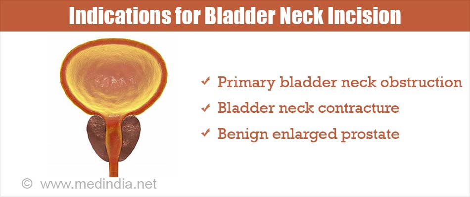
Bladder neck incision is done to facilitate the passage of urine from the urinary bladder. It can be used to treat the following conditions:
Primary bladder neck obstruction: Primary bladder neck obstruction is a functional problem - there is no definite structural cause for the obstruction to the urinary outflow in these cases. The patient may complain of symptoms of reduced urinary stream, dribbling of urine, incomplete emptying of the bladder, urge incontinence, or increased frequency of urination at night.
Bladder neck contracture: Bladder neck contracture is a condition where the bladder neck is narrow due to scar tissue formation. It could occur following prostate surgery, especially if the surgery is followed by radiation therapy through external beam radiation
Benign enlarged prostate: In Some, prostate gland that is small with minimum prostatic enlargement, the incision can help to relieve obstruction to the urinary flow. In these prostates the the capsule of the prostate maybe thick and does not allow the enlarging prostate to expand. The gland itself gets trapped and expands inside the lumen or opening of the urethra and blocks it.

What are the Types of Bladder Neck Incision?
Bladder neck incision may be unilateral or bilateral depending on whether one or two incisions are made on the bladder neck.
The incision may be performed with monopolar or bipolar electrocautery, holmium laser or using a loop resection.
How do you Prepare Before Bladder Neck Incision?
You will have to go through some tests initially to confirm that you will benefit from the procedure. These tests include:
- Uroflometry test: During the uroflowmetry test, the patient is asked to pass urine in a special container, which measures the speed and volume of the urine excreted. After the test the residual urine left behind is also checked. In Bladder neck obstruction the flow is slow and prolonged. There may also be a significant residual urine left after completion of voiding which maybe over 50 mls. Residual urine checking requires an ultrasound probe.
- Videourodynamic tests: These tests study the storage and voiding function of the lower urinary system. During these tests, the bladder is filled with water or contrast and images using x-ray or ultrasound are obtained as the bladder is emptied. Patients with bladder neck obstruction may show an incomplete emptying of the bladder, with some urine remaining in the bladder even after emptying. The pressure of the bladder is generally high. The video test may show that the bladder neck fails to open when the bladder pressure goes up to void.
- Retrograde urethrogram and voiding cystourethrogram: This is done if a bladder neck stricture is suspected after a previous resection of the prostate (TURP or Holmium laser resection). In this test, contrast is introduced into the urinary bladder through the urethra and images are obtained, which may reveal the presence of scar tissue at the bladder neck. In a voiding cystourethrogram, images are obtained when the patient passes out the urine / contrast.
- Electromyography: Electromyography (EMG) refers to the recording of the electrical activity of the muscles of the urinary bladder and the sphincters to detect any functional problems. The sensors are placed on the skin near the urethra or rectum or introduced into the urethra or rectum through catheters. This test is often done in females who need to undergo the bladder neck incision.
Routine tests: Routine tests which are done before any surgery include:
- Blood tests like hemoglobin levels, blood group, and liver and kidney function tests
- Urine tests - Urine infection should be ruled out prior to the procedure through a urine routine and microscopic examination and a urine culture. If present, the infection should be treated prior to the procedure.
- Electrocardiogram (ECG) to study the electrical activity of the heart
- Chest x-ray - In older patients, a detailed assessment of the heart may be required to make sure that they are fit for surgery.
Type of Anesthesia: Bladder neck incision is done under general or spinal anesthesia. If you undergo general anesthesia, you will be asleep during the procedure and will not be aware of what is going on. If you undergo spinal anesthesia, you will not be able to feel any pain waist downwards or move your legs, but will be conscious during the procedure.
Pre-operative Check-up: Routine tests as indicated above are ordered a few days before the surgery. Your doctor may advise you to stop medications like aspirin around a week prior to the procedure. Admission is done on the day of the surgery. Informed consent is obtained. Fasting for around 6 hours before the procedure is usually required.
Shift from the Ward or Room to the Waiting area in the Operating room: An hour or two before the surgery, you will be shifted to the operating room waiting area on a trolley. Once the surgical room is ready, you will be shifted to the operating room.
Anesthesia before Surgery: During general anesthesia, the anesthetist will inject drugs through an intravenous line and make you inhale some gases through a mask that will put you in deep sleep. Once you are asleep, a tube will be inserted into your mouth and windpipe to administer the anesthetic gases to overcome pain and keep you comfortable while the surgery is going on. If you undergo spinal anesthesia, you will receive an injection in your back, following which you will feel numb from your waist downwards. You will be placed in a lithotomy position, with your legs supported on stirrups for the surgery.
During the procedure
First a cystoscope is used to assess the urethra, prostate, bladder neck and the bladder. After this a resectoscope is introduced into the urinary bladder through the urethra. If the bladder neck is extremely narrow and the narrowed part is first dilated using urethral dilators. Then, as the resectoscope is passed in gradually, and incisions are made at predefined positions, usually at the 5- and 7-o’clock position. The incisions extend from just below the ureteric orifices (the openings of the ureters into the urinary bladder) to just above the verumontanum, a bulge noted on the inner aspect of the prostatic urethra in males. The incisions are usually deepened till to cut the capsule of the prostate until the perivesical fat is seen, this is the fat that surrounds the bladder. In males, a small amount of prostate tissue may also be removed.
In males, a modified bladder neck incision is sometimes preferred where the lower one third of the prostatic urethra near the verumontanum is preserved; this approach prevents retrograde ejaculation, that is, backward flow of the semen into the urinary bladder during ejaculation.
Once the procedure is complete, a three way Foley’s catheter to facilitate passage of urine is left in place, and may remain so for around three days following the surgery. Normal saline irrigation maybe used to wash the small blood clots that may form in the bladder from the surgical incision.
After the Procedure:
Waking up from General Anesthesia: Once fully awake after surgery, you will be shifted on the trolley and taken to the recovery room.
Recovery room: In the recovery room, a nurse will monitor your vitals and observe you for an hour or two before shifting you to the room or a ward.
Post-operative recovery:
- You will be encouraged to drink a lot of fluids once you are comfortable.
- Antibiotics will be prescribed to prevent infection and pain relievers to reduce pain if present.
- The urine passed through the Foley’s catheter may contain some blood in the first 12 hours following the surgery, which usually clears up in due course.
- You will be allowed to go home the same day, and will be called back in two to three days for the catheter removal.
- It is important to note that not all your symptoms will disappear overnight but will take some time to settle down.
- Restrict activities for six to eight weeks and follow up with your doctor on a regular basis as advised.
- Pelvic exercises will help in the faster relief of symptoms.
- Sexual activity can be resumed once you are comfortable, may be in around a month after the surgery.
What are the Possible Complications after Bladder Neck Incision?
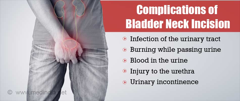
The possible complications of bladder neck incision include the following:
- Burning while passing urine
- Infection of the urinary tract
- Injury to the urethra
- Blood in the urine
- Retrograde ejaculation, which is more common with the bilateral surgery as compared to the unilateral surgery.
- Urinary incontinence due to injury to the external urethral sphincter
- In females - Perforation of the vaginal wall in females
- Requirement for a repeat procedure, and sometimes even an open surgery especially in cases of a cold knife procedure. People who want to avoid a repeat procedure may have to do self-catheterization repeatedly.


