Hysterectomy - Uterus Anatomy
Female Reproductive System
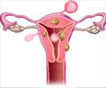
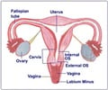
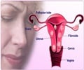
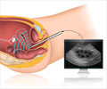
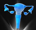
The female reproductive organs are made up of the vulva, the vagina, the uterus, the fallopian tubes, and the ovaries.
The uterus is a hollow, pear-shaped organ that is the home to a developing fetus. The uterus is divided into two parts: the lower part of the uterus that opens into the vagina is called the cervix, and the main body of the uterus, is called the corpus. The uterus has three layers.
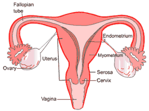
- Endometrium This is the innermost layer of the uterus. The thickness of the endometrium is regulated by hormones and so varies according to the phase of the menstrual cycle. Another name for this lining layer is the mucosa.
- Myometrium This middle layer is a thick wall made of smooth muscle cells.
- Serosa The outermost layer consists of a membrane called the serosa. This thin layer merges with connective tissue (ligaments) that suspends the uterus in the pelvis.
The blood supply to the uterus is through the uterine arteries, which are branches of the Aorta, which is the main blood vessel of the body.
The vagina is a canal that joins the cervix to the outside of the body. It also is known as the birth canal.
The ovaries are small, oval-shaped glands about the size of an almond that are located on either side of the uterus. The ovaries produce eggs and hormones.
The Fallopian tubes are attached to the upper part of the uterus and serve as tunnels for the ova to travel from the ovaries to the uterus. Conception, the fertilization of an egg by a sperm, normally occurs in the fallopian tubes. The fertilized egg then moves to the uterus, where it implants to grow into an embryo.

