- Congenital Mitral Stenosis With or Without Associated Defects - (http://circ.ahajournals.org/cgi/content/full/102/suppl_3/III-166)
- Mitral Stenosis - (http://www.emedicine.com/med/topic1486.htm)
- Hospitalization statistics for Mitral valve disease: - (http://www.wrongdiagnosis.com/m/mitral_valve_disease/stats.htm#medical_stats)
- Congenital Mitral Stenosis - (http://www.emedicine.com/ped/topic2517.htm)
- Mitral valve regurgitation - (http://www.nlm.nih.gov/medlineplus/ency/article/000176.htm)
- Heart & Vascular Institute (Miller Family) - (http://www.clevelandclinic.org/heartcenter/pub/guide/disease/valve/mvrepairfaq.htm)
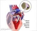
Mitral Valve Replacement
Procedure
Patient will be placed under general anesthesia. An endotracheal tube will be inserted into the patient’s windpipe and connected to a ventilator to assist the patient’s breathing. Another probe will be inserted through the patient’s esophagus. This is a TEE (trans thoracic echocardiogram) probe. TEE allows intra operative evaluation of the valve motion and detection of prosthetic valve insufficiency. It can detect any presence of thrombus as well.
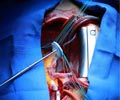
Patient will be cleaned using antiseptic solution like alcoholic spirit and betadine and draped completely except exposing his chest. Routinely the chest is cut along the middle and the breastbone is sawed open. When the heart is exposed, the heart is connected to a heart lung machine. The blood is diverted through sterile plastic tubes to the heart lung machine, which oxygenates and pumps it back to the body. It is easier for the surgeon to operate on a still rather than a beating heart. An incision on the left atrium is made. The mitral valve is exposed and flushed with saline. This allows the surgeon to get a good look at the valve.
The diseased valve along with its subvalvular apparatus is cut. A portion of the posterior valve along with its chordae is left in place to protect the wall of the posterior left ventricle. Calcium deposits must be completely removed from the suturing site in a calcified valve. Calcium bits may not allow proper anchoring of the valve in place or loose calcium bits may travel in blood to cause emboli. The valve site is washed well with saline and suction.
Valve sizers are placed in the annulus to determine the size of the valve required for replacement. The prosthetic valve is then sutured around the mitral valve ring (annulus). The sutures are tied when the valve is seated in place. Saline is flushed through the prosthetic valve to check its competency. Some of the associated defects like atrial septal defect closure or aortic valve replacement can be corrected during this procedure. Before closing the left atrium, absence of air and clots are ensured to prevent thrombosis. Heart lung bypass machine is slowly weaned off. When the heart starts beating on its own again, the prosthetic valve function can be checked using trans thoracic echocardiogram. Drainage tubes will be placed in the chest to drain the excess fluids produced. The chest is then sutured in layers, dressed and bandaged.
Surgeons are currently trying a new less invasive method with newer technology. This procedure takes less time than the conventional method, also less time in the hospital and less recovery time.


Methods for Mitral Valve Repair: Annuloplasty:In annuloplasty an artificial ring is placed around the annulus of the valve.This ring reinforces the annulus and restores the valve to its normal shape and size. Balloon Valvuloplasty:Balloon valvuloplasty is performed using a catheter, i.e. a very thin flexible tube which can be inserted into the body, with a balloon at the end. The balloon is put inside the valve and is expanded thus stretching the valve and bringing it back to its normal size. For more info: heart-consult.com/articles
Various symptoms are
* Shortness of breath.
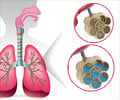
* Pulmonary edema or fluid accumulation in the lungs.
* Orthopnea or shortness of breath while lying flat.
* Paroxysmal nocturnal dyspnea or Cardiac Asthma
* Decreased exercise tolerance.
* Swollen feet or ankles.