What is Photorefractive Keratectomy (PRK)?
Photorefractive keratectomy is a type of laser procedure on the eye to correct refractive errors and presbyopia by reshaping the cornea. After undergoing the procedure, light entering the eye gets properly focused onto the retina for clear vision.It was the first type of laser eye surgery for vision correction and was followed by the laser-assisted-in-situ keratomileusis or the LASIK procedure.
The refractive power of the eye depends primarily on 3 factors
- Curvature of the cornea
- Lens curvature and refractive index
- Antero – posterior diameter (axial length) of the eye
In photorefractive keratectomy, the corneal curvature is altered by reshaping of the cornea using an excimer laser, regardless of the cause of the refractive error, to correct defective vision in patients with refractive errors and presbyopia (age related difficulty in seeing near objects).
The cornea is the transparent part of the front portion of the eye that refracts light entering it. In cross section, it has six microscopic layers – epithelium, Bowman’s membrane, stroma, Dua’s layer, descemet’s membrane and endothelium.
Why is it done?
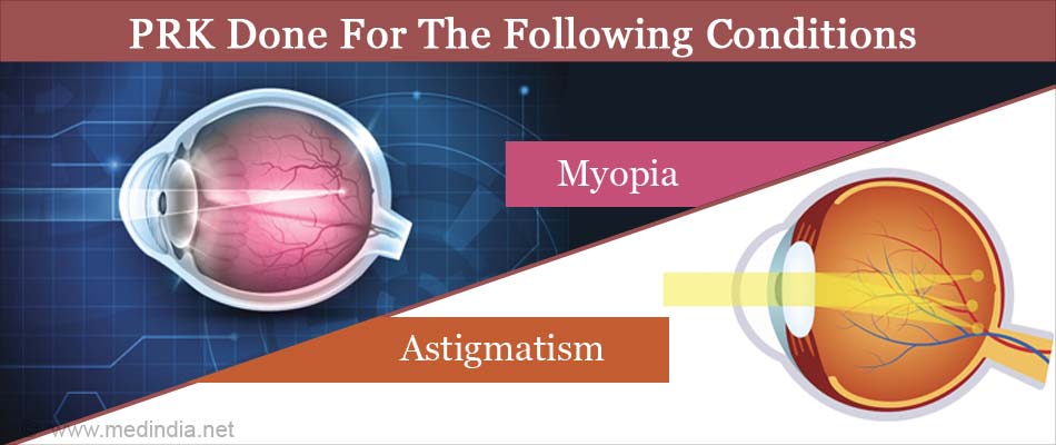
A refractive error results in light rays from an object not falling on the retina after refraction at the cornea and lens. The light rays either fall in front of the retina (myopia), behind the retina (hypermetropia) or differentially in different meridians of the eye (astigmatism). Alternately, in presbyopia (age related physiological weakness of accommodation), images from near objects alone fall behind the retina.
For example, in myopia the rays fall in front of the retina due to excess convergence. Hence the cornea can be flattened to result in a mild divergence of rays (due to reduced converging ability) so that they can fall exactly on the retina. This is done by removal of corneal tissue (ablation) in the center of the cornea. Similarly in hypermetropia, the central portion of the cornea is made more convex to increase convergence by ablating tissue in the form of a ring around the center of the cornea.
In astigmatism, the cornea is shaped differentially according to the refraction.
In presbyopia, either the cornea is converted into a multifocal surface, or one eye is made more convex so that the eye can be used for near vision while the other eye is used for distant vision (monovision).
A 193 nm ultraviolet argon fluoride excimer laser (where excimer stands for excited dimer) is used. Since this type of laser has poor penetrating capacity, it is used for surface ablation (tissue removal) of cornea.

Who can Undergo Photorefractive Keratectomy?
- Patients with low to moderate degrees of short-sightedness or myopia and astigmatism, and low degrees of long-sightedness or hypermetropia
- Patients who have undergone LASIK and who require retreatment
- Patients who cannot undergo LASIK –
- Patients with thin corneas
- Patients where flap stability is critical example individuals in military, law enforcement, and contact sport
What should the Patient Undergoing Photorefractive Keratectomy Expect?
- Photorefractive keratectomy can correct only up to 6 dioptres of myopia, (-6.00 dioptres) low degrees of hypermetropia and 3 dioptres of astigmatism. Higher degrees of refractive errors would require a different procedure such as LASIK or placement of an intraocular lens in the eye.
- Younger patients undergoing refractive procedures should be aware that if abolishment of the refractive error is undertaken, the person would still need to wear glasses by about the age of 40 years due to the onset of presbyopia. Presbyopia affects all near vision tasks such as reading, using mobile phones or computers, for common household chores like stitching or tailoring, looking at a watch or reading a label on a product.
- Patients in the presbyopic age group need to be aware of this aspect, and may consider going in for unilateral correction or under correction if myopic, if they wish to read without glasses after the procedure. A trial of contact lenses prior to the procedure would help the patient understand the situation better, so that they can make an informed decision.
- Photorefractive keratectomy cannot prevent future ocular problems like cataract and glaucoma.
- Also, since photorefractive keratectomy corrects the refractive error at the level of the cornea, and does not affect the retina, any tendency to retinal detachment in people who are prone to it in people with high myopia is not addressed, and the person would still be required to undergo regular ophthalmic screening even if the myopia is corrected.
What is the Difference between PRK and LASIK?
| PRK | LASIK | |
| Depth of cornea at which treatment is performed | Superficial | Deep |
| Corneal flap related complications | No | Yes |
| Pain after the procedure | Can be moderate to severe | Minimal |
| Procedure can be done in thin corneas | Yes | No |
| Risk of ectasia (outward bulging of cornea) after procedure | Less | More |
| Post-operative vision recovery | 3-7 days | Less than 1 day |
| Corneal haze | Quite common | Less common |
When should Photorefractive Keratectomy NOT be done?
- If patient is less than 18 years of age
- If the power is not stable for at least one year
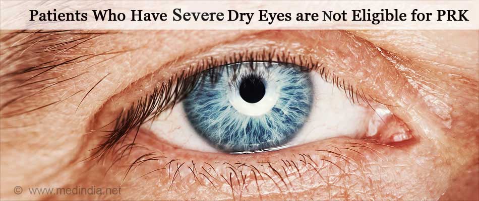
- Severe dry eyes

- Presence of corneal disease
- Presence of other ocular conditions like uncontrolled glaucoma or cataract
- If there is evidence of corneal ectasia (outward bulging) as evidenced on corneal topography, then PRK should not be done as there is a further risk of ectasia after the procedure
- Presence of only one functioning eye
- Pregnancy and lactation
- Immunocompromised states like cancer and HIV / AIDS
- Patients with a tendency for keloid formation
- Diabetics with reduced corneal sensation
- Progressive retinal conditions
PRK should be done with caution in the following conditions:
- Mild dry eyes
- Diminished corneal sensation
- Connective tissue diseases like rheumatoid arthritis, systemic lupus erythematosus, Sjogren syndrome, Wegener’s granulomatosis.
- Any corneal disease
- Patients on medication with isotretinoin, amiodarone, sumatriptan, and topical or oral corticosteroids
What are the Tests Required before Photorefractive Keratectomy?
You will have to be first evaluated by an eye doctor to consider suitability for the procedure
- If you are wearing contact lenses, you will be required to discontinue soft lenses for about 2 weeks and rigid lenses for about 2-3 weeks prior to examination by the eye doctor.
- A complete ophthalmic examination will be carried out which includes testing your visual acuity, refraction, slit lamp examination, measurement of intraocular pressure and examination of your retina.
- Corneal topography (computerised analysis of corneal surface) will be done.
You will be asked to place your chin on the chin rest of the topographer and make your forehead touch the forehead rest. You will see lighted concentric rings. The images of these on your cornea will be used to map the surface configuration of your cornea. This does not hurt and takes just a couple of minutes.
- Pachymetry (measurement of corneal thickness).
The procedure is similar to that described above for corneal topography.
How do you prepare before a PRK Surgery?
- Avoid eye makeup from 3 days prior to surgery
- You may be prescribed antibiotic eye drops from 1-3 days prior to the surgery by your doctor.
- This is a day care procedure and does not require admission.
- You may reach the hospital on the day of your procedure.
What Happens During a PRK Surgery?
Photorefractive Keratectomy is a surface ablation procedure.
- An oral anti-anxiety medication may be recommended to be taken on the morning of the procedure.
- In the operation theatre, you will be made to lie down. The skin around your eyes will be cleaned and sterilized using povidone – iodine or alcohol.
- A sterile drape will be placed over your face, and local anesthetic eye drops will then be instilled in your eyes to numb any pain. The entire procedure is painless.
- An instrument will then be placed in your eye to keep the eyelids separated. You might feel some pressure but there will be no pain.The epithelium of the cornea is removed either by a blade, absolute alcohol or excimer laser.
- Tissue ablation of bowman’s membrane and superficial stroma is carried out. The tissue removal is achieved by destruction of intermolecular bonds. The location of ablation is determined by the type of refractive condition – central ablation for short-sightedness or myopia, and mid-peripheral ablation for long-sightedness or hypermetropia.
- The laser machine is placed some distance above the eye. You will be asked to have a fixed gaze towards the center of the light of the machine.
- The laser is centered and focused and laser energy is delivered to the eye to reshape the corneal tissue
- In some cases, to reduce the chance of post-procedure corneal haze, a sponge soaked in M mitomycin C is placed on the treated surface for about 12 seconds.
- A bandage contact lens (a soft contact lens to promote corneal epithelial healing) will then be placed on the eye
- The entire procedure takes about 10-15 minutes.
- You will have to wait in the recovery room for about half an hour, after which you can go home.
What Happens after a PRK Surgery?
Post-operative recovery:
- Pain starts about half an hour after the procedure and lasts for a few days. The pain may be alleviated by anti-inflammatory eye drops. The bandage contact lenses also play a major part in pain management.
- You will be instructed to instill antibiotic eye drops, corticosteroid eye drops, and sometimes non-steroidal anti-inflammatory eye drops.
- As soon as the epithelium is healed (3-5 days) the bandage contact lens is removed and antibiotic eye drops and non-steroidal anti-inflammatory eye drops are discontinued. Corticosteroid eye drops may be continued for a variable period of time, which will be decided by the eye doctor.
- Corneal haze following photorefractive keratectomy is almost universal, usually occurs within the first month and lasts a couple of months. It usually does not interfere with vision.
- Dryness of eyes is usually present for a few months after the procedure. Discomfort due to dryness may be addressed by the use of artificial tears.
- Vision after photorefractive keratectomy takes a couple of days to settle down, and may sometimes take a couple of months to stabilize.
- Reduction of low contrast visual acuity and night vision symptoms like glare and halos may occur for a couple of months following photorefractive keratectomy. This can cause difficulty in driving at night for some people.
- Instructions following discharge.
- You should not drive for a couple of days after the procedure, and should wear sunglasses when outside.
- Avoid getting sweat and water inside the eyes for about 2 weeks. Hence exercise is best avoided for 2 weeks.
- You may bathe the next day, but water should not get into the eyes.
- The eyes should not be rubbed.
- Eye makeup and creams should not be used for one week.

What are the Complications and Risks of PRK Surgery?
PRK is a relatively safe surgery. However sometimes complications can occur as follows:
- Overcorrection or under-correction
- Optical aberrations
- De-centered ablations
- Dry eyes and diminished corneal sensation
- Infection of the cornea
- Persistent corneal haze
- Scarring of cornea
- Raised intraocular pressure due to corticosteroid use


