- Feusner JD, Madsen S, Moody TD, Bohon C, Hembacher E, Bookheimer SY, et al. Effects of cranial electrotherapy stimulation on resting state brain activity. Brain Behav. 2012 May; 2(3): 211-20. PMCID: PMC3381625. DOI - (10.1002/brb3.45)
- Ling LU, Jun HU. A Comparative study of anxiety disorders treatment with Paroxetine in combination with cranial electrotherapy stimulation therapy. Medical Innovation of China 2014;11(08): 080-082. - (10.1002/brb3.45)
- Barclay TH, Barclay RD. A clinical trial of cranial electrotherapy stimulation for anxiety and comorbid depression. J Affect Disord. 2014; 164: 171-7. DOI - (10.1016/j.jad.2014.04.029)
- Kirsch DL, Nichols F. Cranial electrotherapy stimulation for treatment of anxiety, depression, and insomnia. Psychiatr Clin N Am. 2013; 36(1): 169-76. DOI - (10.1016/j.psc.2013.01.006)
- Rosa MA, Lisanby SH. Somatic treatments for mood disorders. Neuropsychopharmacology 2012; 37: 102-16. DOI - (10.1038/npp.2011.225)
- Koleoso ON, Osinowo HO, Akhigbe KO. The role of relaxation therapy and cranial electrotherapy stimulation in the management of dental anxiety in Nigeria. IOSR Journal of Dental and Medical Sciences 2013; 10(4): 51-7. - (10.1016/j.jad.2014.04.029)
- Kavirajan HC, Lueck K, Chuang K. Alternating current cranial electrotherapy stimulation (CES) for depression. Cochrane Database of Systematic Reviews 2014 - (10.1002/14651858.CD010521.pub2)
What is Cranial Electrotherapy Stimulation?
Cranial electrotherapy stimulation (CES) involves cranial stimulation with pulsed, low-intensity current, usually applied to the earlobes or over the mastoid process or the maxilla-occipital junction.
The CES Device: CES uses a medical device about the size of a cell phone. The device is a nerve stimulator and is capable of sending a pulse of weak electrical current to the brain through electrodes. The device produces a current that cannot be sensed by the wearer (usually below 4 milliamps), preset to 0.5 Hz, and emitted in cycles for 20-minute countdown to auto-off. The CES device is recognized by the U.S. Food and Drug Administration (USFDA) as a Class III device as a prescriptive, non-invasive, electro-medical treatment for insomnia, depression, and anxiety.
Anatomy & Physiology of the Cranium in Brief
The human skull consists of 22 bones which are mostly connected together by ossified joints, so called sutures. The skull is divided into the brain case (cerebral cranium) and the face (viscero-cranium). There are 8 cranial bones and 14 facial bones, adding up to a total of 22 bones. The brain case houses the brain. The face consists of the facial bones that form the upper and lower jaws, nose, orbits, and other facial structures. The major function of the cranium is the protection of the most important organ in the human body, namely, the brain.
What is the Purpose of the Cranial Electrotherapy Stimulation Therapy?
CES, also known as transcranial electrostimulation is one of the first techniques that was developed for stimulating the brain non-invasively. It encompasses several different techniques that use low-level alternating current (AC) applied to the scalp or earlobes
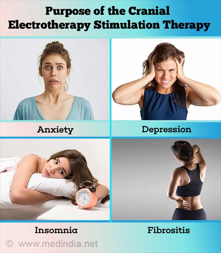
- The main purpose of CES is for the treatment of anxiety, depression and insomnia.
- It has also been used for the treatment of pain, headaches, quitting smoking, fibrositis, and giving-up drugs.

However, a 2014 Cochrane review found insufficient evidence to determine whether or not CES using AC is safe and effective for treating depression.
Mechanism of Action of CES
The mechanism of action of CES is not clearly understood.
- Available evidence suggests that feeble AC applied to the cranium of a sleeping patient can affect memory and fluctuations in brain activity.
- The levels of neurotransmitters and hormones can be altered. For example, it has been found that thyroxine production is increased as a result of brain stimulation. Studies have indicated CES elevated blood plasma levels of endorphins, adrenocorticotrophic hormone, serotonin, melatonin, norepinephrine, and cholinesterase. CES has also been shown to decrease serum cortisol levels
- Increase in platelet monoamine oxidase-B (MAO-B) activity and plasma gamma-amino butyric acid (GABA) concentrations have also been observed.
- Moreover, changes in electroencephalogram (EEG) readings during and after stimulation have been reported, especially slowing of α waves.
However, due to the lack of animal studies, there is a possibility that the effects may be mediated via cranial nerve stimulation rather than direct brain stimulation.
How is it Performed?
CES is performed quite easily since there are several user-friendly handheld devices available in the market. Nowadays, CES can be performed independently by the patients themselves, without any help. Depending of the type of CES stimulator, the electrodes are placed on the ear lobes, maxilla-occipital junction, mastoid processes or temples. The most popular and user-friendly site for attachment of the electrodes is the ear lobe. The pre-programmed stimulation parameters are set in the handset as per requirement and a switch is pressed to start the stimulation. The entire stimulation process is fully automated.
It has been found that CES stimulation at a current strength of 1 mA (milliampere) is capable of sending pulses to the thalamus. As a result, changes are induced in the EEG pattern, with increased frequency of α waves and decreased frequencies of δ and β waves, which are characteristic brain waves that are observed in various states of brain activity.
Stimulator Models: Currently available CES stimulators are listed below:
- Fisher–Wallace Cranial Stimulator (Model SBL500-B)
- Liss Cranial Stimulator (Model SBL201-M)
- Alpha-Stim® Stress Control System
Side Effects of CES: Headache, vertigo and nausea are the most common side effects of CES, followed by skin irritation at the site where the electrodes are attached.


