The first to show, in mice, an unexpected two-step process that happens during the growth and regeneration of inner ear tip links is a team of NIH-supported researchers.
The findings appear in the June 11, 2013 online edition of PLOS Biology. The senior author of this study is Gregory I. Frolenkov, an associate professor in the College of Medicine at the University of Kentucky, Lexington, and his fellow, Artur A. Indzhykulian, Ph.D., is the lead author.
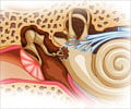
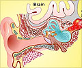
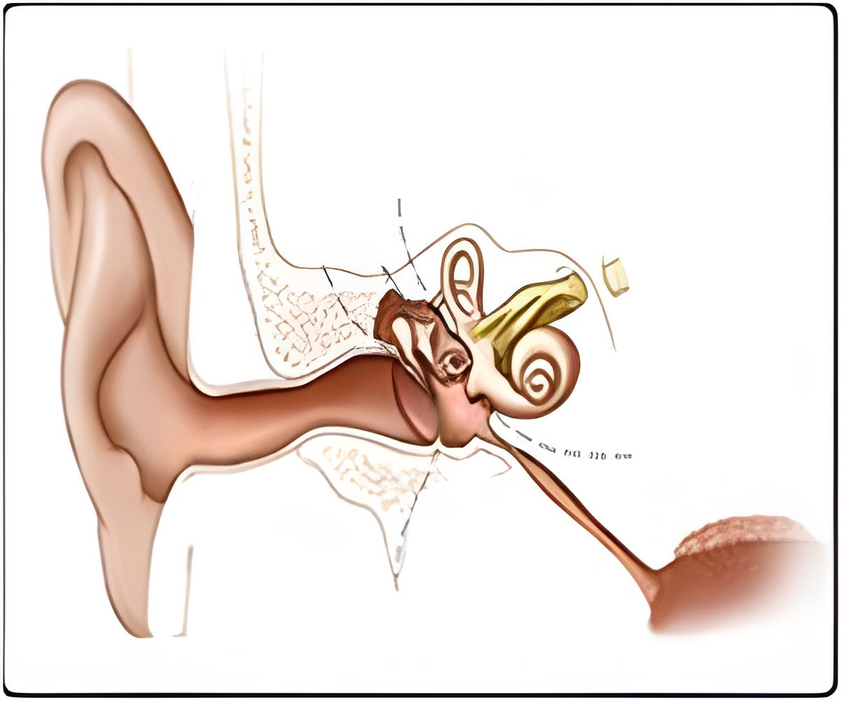
Stereocilia are bundles of bristly projections that extend from the tops of sensory cells, called hair cells, in the inner ear. Each stereocilia bundle is arranged in three neat rows that rise from lowest to highest like stair steps. Tip links are tiny thread-like strands that link the tip of a shorter stereocilium to the side of the taller one behind it. When sound vibrations enter the inner ear, the stereocilia, connected by the tip links, all lean to the same side and open special channels, called mechanotransduction channels. These pore-like openings allow potassium and calcium ions to enter the hair cell and kick off an electrical signal that eventually travels to the brain where it is interpreted as sound.
The findings build on a number of recent discoveries in laboratories at NIDCD and elsewhere that have carefully plotted the structure and function of tip links and the proteins that comprise them. Earlier studies had shown that tip links are made up of two proteins -- cadherin-23 (CDH23) and protocadherin-15 (PCDH15) – that join to make the link, with PCDH15 at the bottom of the tip link at the site of the mechanotransduction channel, and CDH23 on the upper end. Scientists assumed that the assembly was static and stable once the two proteins bonded.
Tip links break easily with exposure to noise. But unlike hair cells, which can't regenerate in humans, tip links repair themselves, mostly within a matter of hours. The breaking of tip links, and their regeneration, has been known about for many years, and is seen as one of the causes of the temporary hearing loss you might experience after a loud blast of sound (or a loud concert). Once the tip links regenerate, hair cell function returns usually to normal levels. What scientists didn't know was how the tip link reassembled.
To study tip link assembly, the researchers treated young, postnatal (5-7 days) mouse sensory hair cells with BAPTA – a substance that, like loud noise, damages and disrupts tip links. To image the proteins, the group pioneered an improved scanning electron microscopy (SEM) technique of immunogold labeling that uses antibodies bound to gold particles that attach to the proteins. Then using SEM they imaged the cells at high resolution to determine the positions of the proteins before, during, and after BAPTA treatment.
Advertisement
Why tip links regenerate using a two-step instead of a neat one-step process is not known. For reasons that are still unclear, CDH23 disappears from stereocilia after noise damage while PDCH15 stays around. Looking to regenerate quickly, the lower PDCH15 latches onto another PDCH15, forming a shorter and functionally slightly weaker tip link. Later, at some time during the 36 hours after the damage, when CDH23 returns, PDCH15 gives up its provisional partner and latches onto its much stronger mate in CDH23. In other words, PDCH15 prefers to be with CDH23, but in a pinch it will bond weakly with another bit of PDCH15 until CDH23 shows up.
Advertisement
Additional experiments revealed that when hair cells develop, the tip links use the same two-step process.
Previous research has shown that both CDH23 and PCDH15 are required for normal hearing and vision. In fact, NIDCD scientists in earlier studies have shown that mutations in either of these genes can cause the hearing loss or deaf-blindness found in Usher Syndrome types 1D and 1F.
"In the case of deaf individuals who are unable to make functional CDH23, knowledge of this new temporary alliance of PCDH15 proteins to form a weaker, but still functional, tip link could inform treatments that would encourage the double PCDH15 bond to become permanent and maintain at least limited hearing," said Tom Friedman, Ph.D., chief of the Laboratory of Molecular Genetics at NIDCD where the research began.
Source-Eurekalert


