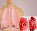
A lung transplant is currently the only cure for idiopathic pulmonary fibrosis, a condition that causes scarring in the lungs.

‘Organoids, created with human pluripotent or genome-edited embryonic stem cells, may be the best, and perhaps only, way to gain insight into the pathogenesis of the disease.’


By reproducing an organ in a dish, researchers hope to develop better models of human diseases, and find new ways of testing drugs and regenerating damaged tissue. "Researchers have taken up the challenge of creating organoids to help us understand and treat a variety of diseases," said lead investigator of the study Hans-Willem Snoeck, Professor at Columbia University Medical Centre in the US.
"But we have been tested by our limited ability to create organoids that can replicate key features of human disease," Snoeck said.
The lung organoids created in Snoeck's laboratory, however, include branching airway and alveolar structures, similar to human lungs, according to a study published in the journal Nature Cell Biology.
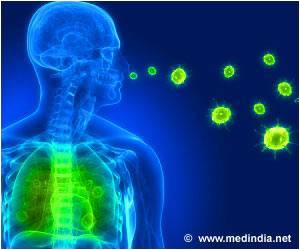
To demonstrate their functionality, the researchers showed that the organoids reacted in much the same way as a real lung does when infected with respiratory syncytial virus (RSV).
Advertisement
Respiratory syncytial virus is a major cause of lower respiratory tract infection in infants and has no vaccine or effective antiviral therapy.
Advertisement
"Organoids, created with human pluripotent or genome-edited embryonic stem cells, may be the best, and perhaps only, way to gain insight into the pathogenesis of these diseases," Snoeck said.
Source-IANS

