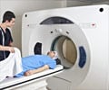
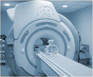
According to a study among six large integrated health care systems between 1996 and 2010 there was a substantial increase in the use of advanced diagnostic imaging.

"Most studies that have evaluated patterns of diagnostic imaging have assessed insurance claims for fee-for-service insured populations where financial incentives encourage imaging. No large, multisite studies have assessed imaging trends in integrated health care delivery systems that are clinically and fiscally accountable for the outcomes and health status of the population served. Understanding imaging utilization and associated radiation exposure in these settings could help us determine how much of the increase in imaging may be independent of direct financial incentives," the researchers write.
Rebecca Smith-Bindman, M.D., of the University of California, San Francisco, and colleagues conducted a study to estimate trends in imaging utilization and associated radiation exposure among members of integrated health care systems. The study consisted of an analysis of electronic records of members of 6 large integrated health systems from different regions of the United States. Review of medical records allowed estimation of radiation exposure from selected tests. Between 1 million and 2 million member-patients were included each year from 1996 to 2010. Enrollees underwent a total of 30.9 million imaging examinations during the study period, reflecting an average of 1.18 tests per person per year, of which 35 percent involved advanced diagnostic imaging (i.e., CT, MRI, nuclear medicine, and ultrasound).
The researchers found that use of radiography and angiography/fluoroscopy rates were relatively stable over time: radiography increased 1.2 percent per year, and angiography/fluoroscopy decreased 1.3 percent per year. "In contrast, the utilization of advanced diagnostic imaging changed markedly. Computed tomography examinations tripled (52/1000 enrollees in 1996 to 149/1000 in 2010, 7.8 percent annual growth); MRIs quadrupled (17/1000 to 65/1000,10 percent annual growth); ultrasounds approximately doubled over the same period (134/1000 to 230/1000, 3.9 percent annual growth). Nuclear medicine rates decreased (32/1000 to 21/1000, 3 percent annual decline), although after 2004, PET imaging rates increased from 0.24 per 1,000 enrollees to 3.6 per 1,000 enrollees, 57 percent annual growth."
The authors also found that the increase in the utilization of CT was associated with an increase in estimated exposure to radiation, with the average per capita effective dose increasing from 1.2 mSv in 1996 to 2.3 mSv in 2010. The percent of enrollees who received high (> 20-30 mSv) or very high (> 50 mSv) radiation exposure during a given year also approximately doubled across study years. The researchers also estimated that by 2010, 2.5 percent of enrollees received a high annual dose of greater than 20 to 50 mSv, and 1.4 percent received a very high annual dose of greater than 50 mSv. By 2010, 6.8 percent of patients who underwent imaging received a high dose of more than 20 to 50 mSv and 3.9 percent of patients received a very high dose above 50 mSv during this single year.
The researchers write that the "increase in imaging use over this period was likely driven by many factors, including improvements in the technology that have led to expansion of clinical applications, patient- and physician-generated demand, defensive medical practices, and medical uncertainly—all factors that would be expected to influence utilization across all systems of medical care."
Advertisement
Source-Eurekalert


