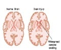
A study found that patients with traumatic brain injury (TBI) had increased deposits of Amyloid plaques in some areas of their brains.

Researchers used imaging and brain tissue acquired during autopsies to examine Aβ deposition in patients with TBI. Researchers performed positron emission tomography (PET) imaging using carbon 11-labeled Pittsburgh Compound B ([ 11 C]PIB), a marker of brain amyloid deposition, in 15 participants with a TBI and 11 healthy patients. Autopsy-acquired brain tissue was obtained from 16 people who had a TBI, as well as seven patients with a nonneurological cause of death.
The study's findings indicate that patients with TBI showed increases in [ 11 C]PIB binding, which may be a marker of Aβ plaque in some areas of the brain.
"The use of ([ 11 C]PIB PET for amyloid imaging following TBI provides us with the potential for understanding the pathophysiology of TBI, for characterizing the mechanistic drivers of disease progression or suboptimal recovery in the subacute phase of TBI, for identifying patients at high risk of accelerated AD, and for evaluating the potential of antiamyloid therapies," the authors conclude.
Source-Eurekalert


