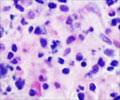
Analyzing tumor DNA fragments in the bloodstream may give a more complete picture of the molecular basis of a patient's cancer.

‘The molecular mechanism of resistance that emerges in one metastatic lesion while on a given therapy can be totally different from the mechanism driving resistance in a neighboring lesion in the same patient.’


"We found that the molecular mechanism of resistance that emerges in one metastatic lesion while on a given therapy can be totally different from the mechanism driving resistance in a neighboring lesion in the same patient," says Ryan Corcoran, MD, PhD, translational research director for the Center for Gastrointestinal Cancers at the MGH Cancer Center, co-corresponding author of the report. "Our conclusion is that the standard practice of performing molecular testing on a biopsy from a single metastatic lesion may be inadequate and that circulating tumor DNA testing may better capture the molecular diversity in a patient's tumor, enhancing our ability to select effective therapies to overcome resistance."
The authors report the case of a patient with colorectal cancer metastatic to the liver, who responded to an EGFR antibody drug - cetuximab (Erbitux) - for 15 months. When the patient's disease became resistant to therapy, a biopsy of a single liver metastasis was performed to analyze for new, resistance-conferring mutations.
The specimen was found to have an MEK1 mutation that had not been present before cetuximab treatment, and the authors demonstrated that MEK1 mutation could drive resistance to EGFR antibodies in laboratory models of colorectal cancer. Based on those findings, the patient was treated with a combination of the MEK inhibitor trametinib (Mekinist) and another EGFR antibody panitumumab (Vectibix) - a combination that had been effective in a cellular model of cetuximab-resistant tumor cells with the same mutation.
While the patient initially appeared to respond to combination therapy, an abdominal CT scan three months later revealed that, while the MEK1-mutated liver lesion was smaller, additional metastatic lesions had continued growing.
Advertisement
While that KRAS mutation was found nowhere in the MEK1-mutated lesion, it was detected in one of the lesions that continued progressing during combination therapy, implying that separate resistance mutations had developed and were driving the growth of different metastases. Noting the ability of circulating tumor DNA analysis to identify both resistance mutations, the authors emphasize that such "liquid biopsies," which can be repeated frequently and with little inconvenience to the patient, may be better for treatment monitoring than biopsies of single lesions.
He adds, "There has been dramatic progress in our ability to analyze circulating tumor DNA in recent years, and the technology continues to evolve. But while some assays are currently available for clinical use, we are not at a point where these liquid biopsies can replace tumor biopsies entirely. We need to learn how best to exploit the potential of liquid biopsies to provide real-time monitoring of a patient's tumor as it develops resistance to therapy, allowing us to anticipate both the timing and the cause of resistance and modify treatment accordingly."
Source-Eurekalert


