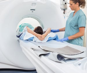
An MRI technique called Diffusion Tensor Imaging (DTI) can now predict cancer outcomes and their response to immunotherapy treatment.

‘The higher the level of reactive immune cells around tumors the longer a patient survives, irrespective of the cancer type or other biological parameters. These reactive cells can be detected by an MRI technique called Diffusion Tensor Imaging.’


The major problem hindering the successful treatment of commonly-occurring cancers is not the primary tumor, which can usually be removed by surgery, but its spread or ’metastasis’ to other organs in the body, forming secondary tumors. One of the most frequent sites of metastasis is the brain. Secondary brain tumors may also reflect the presence of further secondaries elsewhere in the body, any one of which can lead to the death of the patient.
As a general rule, cancer that has spread is treated with chemotherapy or with targeted therapies such as immunotherapy - a relatively new treatment that works by stimulating the body’s immune system to fight cancer.
Immunotherapy is revolutionizing the way doctors treat cancer as it does not come with many of the debilitating side effects produced by chemotherapy. However, it does not work for everyone or every type of cancer, and although successful in some cases, there is currently no simple test to determine who is likely to benefit.
To investigate why some patients with secondary brain cancer do better than others, researchers at the University of Liverpool’s Department of Biochemistry and The Walton Centre Neurosurgery Department used an MRI technique called Diffusion Tensor Imaging (DTI) to analyse brain tumors from appropriate patients and then to sample the same areas for comparative biochemical tests.
Advertisement
The project was led by biochemist Professor Philip Rudland at the University of Liverpool and consultant neurosurgeon Michael Jenkinson at The Walton Centre, Liverpool, with Neurosurgical Registrar and Research Fellow Dr. Rasheed Zakaria bridging the gap between the two teams.
Dr. Zakaria added: "There is huge excitement about immune therapy in brain tumors, but without the time and risk of a neurosurgical operation it is impossible to know which patients should have treatment. Since most patients with a brain tumor undergo MRI scanning we asked the simple question, are there any changes on the MRI scans that correlate with the brain’s immunological reaction to cancer?"
Mr. Jenkinson commented: "The next steps are to repeat this study in a larger group of patients with brain metastasis to validate the findings and then to trial this approach in selecting patients for immunotherapy."
The research draws upon material in the Walton Research Tissue Bank, which provides researchers with access to brain tumor tissue and blood samples to help facilitate the development and testing of new treatments.
Source-Eurekalert


