A simple magnetic resonance imaging (MRI) test involving breathing oxygen might help oncologists determine the best treatment for some cancer patients
Researchers at UT Southwestern Medical Center have revealed that a simple magnetic resonance imaging (MRI) test involving breathing oxygen might help oncologists determine the best treatment for some cancer patients.
Prior research has shown that the amount of oxygen present in a tumor can be a predictor of how well a patient will respond to treatment. Tumors with little oxygen tend to grow stronger and resist both radiotherapy and chemotherapy. Until now, however, the only way to gauge the oxygen level in a tumor, and thus determine which treatment might be more effective, was to insert a huge needle directly into the cancerous tumor.The new technique, known as BOLD (blood oxygen level dependent) MRI, can detect oxygen levels in tumors without the need for an invasive procedure. The patient need only be able to breathe in oxygen when undergoing an MRI.
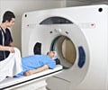
"The patient simply inhales pure oxygen, which then circulates through the bloodstream, including to the tumors," said Dr. Ralph Mason, professor of radiology, director of the UT Southwestern Cancer Imaging Center and senior author of a study appearing online and in a future edition of Magnetic Resonance in Medicine. "Using MRI, we can then go in and estimate how much oxygen a particular tumor is taking up, providing us some insight into how the tumor is behaving and what sort of treatment might be effective."
The most important finding, Dr. Mason said, is that BOLD MRI performed as well as the standard yet more invasive procedure for viewing tumors. That method, known as FREDOM (fluorocarbon relaxometry using echo planar imaging for dynamic oxygen mapping) MRI, requires the injection of a chemical called a reporter molecule directly into the tumor.
"The BOLD technique appears to indicate accurately the oxygen levels in tumors," Dr. Mason said. "Because BOLD is immediately applicable to patients, this holds promise as a new method for predicting response to therapy."
BOLD MRI has been used extensively in studying brain function, but the procedure has only recently begun to be used to assess blood oxygenation and vascular function in tumors.
Advertisement
In the published study, researchers took multiple images of breast tumors implanted just below the skin of rats, which were given anesthesia to help them remain still during the imaging process. Humans do not require anesthesia, Dr. Mason said.
Advertisement
Dr. Mason said that examining each form of cancer in this way presents its own technical challenges. For example, the motion of the lungs or the design of a face mask for breathing oxygen must be taken into account.
"If we can prove that the test is meaningful in animals, then it is that much more worthwhile to argue for doing it in a patient. This preclinical work provides the foundation for future clinical studies," Dr. Mason said. "It helps justify doing a larger clinical trial with the goal of ultimately becoming a diagnostic test for oncologists."
Researchers currently are trying to determine how much oxygen must be inhaled by a patient in order to be effective. The next step is to expand studies in patients and prove the relevance to more tumor types.
Source-Eurekalert
RAS


