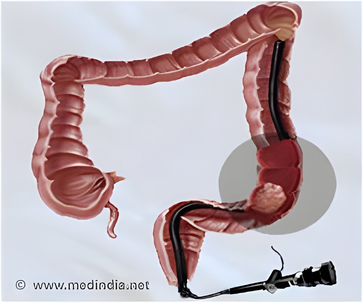
Colonoscopy or flexible sigmoidoscopy can be used to predict Parkinson's disease, say scientists.

A protein called alpha-synuclein is deposited in cells of the brain of patients with Parkinson's disease and is considered a pathologic hallmark of the disorder. These protein aggregates form Lewy bodies, a characteristic structure seen in Parkinson's disease brains at autopsy. Identification of the role of alpha-synuclein aggregation in neuronal dysfunction and death has broadened understanding of how Parkinson's disease develops and introduced a valuable tool for tracking its progress.
Physicians at Rush have demonstrated that the alpha-synuclein protein can also be seen in the nerve cells in the wall of the intestines in research subjects with early Parkinson's disease, but not in healthy subjects. In this study, 10 subjects with early Parkinson's disease had flexible sigmoidoscopy, a technique like colonoscopy, in which a flexible scope is inserted into the lower intestine. In the flexible sigmoidoscopy technique, the scope is only inserted about 8 inches and no colon preparation or anesthesia are required. The procedure takes only 5-10 minutes.
Now, a group of Rush scientists has become the first to demonstrate alpha-synuclein aggregation in biological tissue obtained before onset of motor symptoms of Parkinson's disease.
The studies, published the May 15 issue of the journal Movement Disorders, were conducted by Dr. Kathleen M. Shannon, neurologist in the Movement Disorders and Parkinson's Center at Rush, and a multidisciplinary team of scientists at Rush. The team analyzed samples of tissue obtained during colonscopy examinations that took place 2-5 years before the first symptom of Parkinson's disease appeared in 3 research subjects, and all 3 showed the characteristic protein in the wall of the lower intestine.
"Recent clinical and pathological evidence supports the notion that Parkinson's disease may begin in the intestinal wall then spread through the nerves to the brain. Clinical signs of intestinal disease, such as constipation, Parkinson's disease diagnosis by more than a decade. These studies suggest it may one day be possible to use colonic tissue biopsy to predict who will develop motor Parkinson's disease," said Shannon.
Advertisement
Alternatively, the Rush investigators showed that colonic tissue is easily obtained using flexible sigmoidoscopy, a technique that, unlike colonoscopy, requires no colon cleansing preparation or sedation, and can be performed in 10 minutes.
Advertisement
"In view of a multi-billion-dollar translational research effort that aims to identify agents that slow or stop the progression of Parkinson's disease, the need for accurate and timely diagnostic biomarkers, including the potential for pre-motor diagnosis, is particularly acute," the authors stated.
"We believe that alpha-synuclein in the colonic submucosa may be a pre-motor biomarker that easily can be studied in cohorts at increased risk of developing Parkinson's disease (relatives of Parkinson's disease subjects, subjects with anosmia [inability to smell], rapid eye movement sleep behavior disorder and others)."
The Rush scientists stressed the need to replicate this finding in other populations, including normal controls as well as in subjects with other neurodegenerative Parkinson's-like disorders, and to determine the safest and highest-yield biomarker site.
Source-Eurekalert


