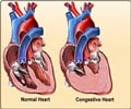
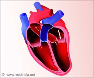
Researchers are collaborating to create a new computer model of the intricate structures of the human heart as part of a larger effort to develop a potentially life-saving heart surgery.

Within the human heart are numerous small muscle bundles called the trabeculae carneae. Despite their significance to the heart’s anatomy, their function is not well understood, and most models of the heart ignore them.
As people grow older, heart muscles can grow stiff, reducing efficiency and sometimes resulting in untreatable diastolic heart failure. SwRI and UTSA are scanning cadaver hearts using a powerful computer tomography (CT) scanner at SwRI to inform a potential new surgical intervention.
“Capturing the intricate structures of the trabeculae carneae requires something more powerful than an MRI or standard CT scanners,” Bartels said. “We’ll utilize a micro-focus X-ray CT scanner here at SwRI to create images of explanted human hearts.”
The images of the heart’s intricate inner structures will help Han to create a realistic anatomical model of the trabeculae carneae, building on a previous model he developed for the left ventricle.
“This collaboration with SwRI is a first step toward creating a new surgical method,” Han explained. “The computer model will help to provide a much deeper understanding of the trabeculae carneae.”
Advertisement
“I hope that this work can ultimately improve the quality of people’s lives and even save lives in the long run,” Bartels said. “Heart failure is a major problem that affects millions of people.”
Advertisement
Source-Eurekalert


