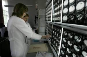
A new study has found that PET/CT scanning is helpful in detecting breast tumours which have reached nearby bones.

Bone scans detect bony regions in the process of growth or repair, which can be a sign of metastatic disease. PET, on the other hand, assesses irregularities of biochemical activity in the body, such as cells that metabolise glucose unusually fast. Such behaviour is a trademark of a cancer cell. Meanwhile, CT creates anatomical images that can help physicians isolate and analyse the size and shape of tumours.
"In contrast with bone scans, which are only able to detect bone metastases, PET/CT has the advantage of concurrently imaging other common sites of breast cancer metastases such as the liver and lungs," said lead author Patrick Morris, a breast cancer specialist at Memorial Sloan-Kettering.
Morris added that the results should be interpreted with caution.
"If this data holds true in a prospective trial, the issue can be laid to rest and PET/CT could replace CT plus bone scan," noted Maxine Jochelson, Director of Radiology for the Breast and Imaging Center.
The study is published in the June 2010 issue of the Journal of Clinical Oncology.
Advertisement


