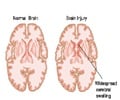
Surgeons find it extremely hard to have a long-term electrode implanted in the brain. But drilling a small hole into the skull could be an effective way out.
Surgeons find it extremely hard to have a long-term electrode implanted in the brain. But drilling a small hole into the skull could be an effective way out, say researchers from the University of California at Berkeley.
Electroencephalography (EEG) records brain activity from electrodes on the skull, but the signal gets 'smeared' as the electrical charge passes through the bone and so the source of the activity can't be located very precisely to specific brain areas. Now a way has been found to obtain better signals - through an operation involving the removal of a portion of the skull, called hemicraniectomy.It is an operation doctors resort to when treating patients who have suffered severe head trauma, such as gunshot or knife wounds. The surgeon cuts out a chunk of skull that’s the diameter of an orange or grapefruit, to give the brain room to swell.
The piece of bone is usually reattached four to six months later, once the swelling has subsided and the skin has healed. In the meantime, the patient’s scalp and a helmet protect the exposed area. And doctors stitch the skull fragment into the abdomen, “bathed in the body’s own fluids,” to prevent it from deteriorating.
The Berkley team took advantage of this brief window of time to compare EEG signals from people with and without the skull as a barrier. Patients performed simple tasks like squeezing a person’s hand or listening to an “oddball stimulus” of three low-pitched sounds followed by a higher one.
During these tasks the team measured a patient’s brain waves on both sides of his head. On one side, just a thin flap of skin separated the brain from the EEG electrode, while on the other side the skull was intact. Signals from the skull-free side were, unsurprisingly, much stronger, less noisy and easier to pinpoint to a specific task and region of the brain.
"Human electrophysiological research is generally restricted to scalp EEG, magneto-encephalography, and intracranial electrophysiology. Here we examine a unique patient cohort that has undergone decompressive hemicraniectomy, a surgical procedure wherein a portion of the calvaria is removed for several months during which time the scalp overlies the brain without intervening bone. We quantify the differences in signals between electrodes over areas with no underlying skull and scalp EEG electrodes over the intact skull in the same subjects," the researchers said in their study published in the Journal of Cognitive Neuroscience.
Advertisement
“If someone’s had a stroke or they’re paralyzed, in the future, the goal of the surgeon is to be able to implant the electrodes into the person’s brain,” he said.
Advertisement
Source-Medindia
GPL


