Promising results for earlier diagnosis of Hepatocellular carcinoma, which is the most common cancer to strike the liver, have been obtained by a research team
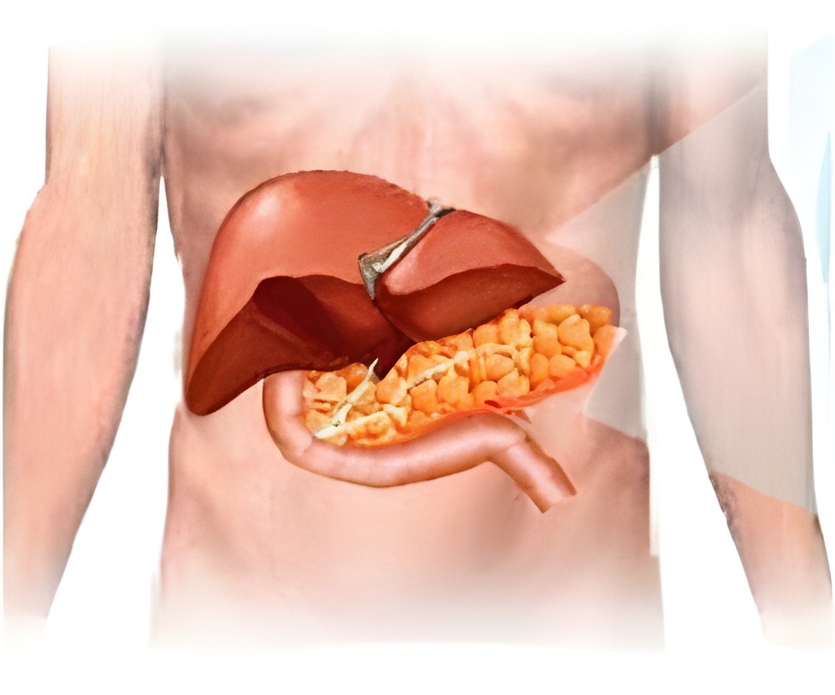
It is the first time that metal nanoparticles have been used as agents to enhance X-ray scattering signals to image tumour-like masses.
"What we're doing is not a screening method," said Christoph Rose-Petruck, professor of chemistry at Brown University and corresponding author on the paper.
"But in a routine exam, with people who have risk factors, such as certain types of hepatitis, we can use this technique to see a tumor that is just a few millimeters in diameter, which, in terms of size, is a factor of 10 smaller," added Rose-Petruck.
The team took gold nanoparticles of 10 and 50 nanometers in diameter and ringed them with a pair of 1-nanometer polyelectrolyte coatings. The coating gave the nanoparticles a charge, which increased the chances that they would be engulfed by the cancerous cells. Once engulfed, the team used X-ray scatter imaging to detect the gold nanoparticles within the malignant cells.
In lab tests, the nontoxic gold nanoparticles made up just 0.0006 percent of the cell's volume, yet the nanoparticles had enough critical mass to be detected by the X-ray scatter imaging device.
Advertisement
The study has been detailed in the American Chemical Society journal Nano Letters.
Advertisement

