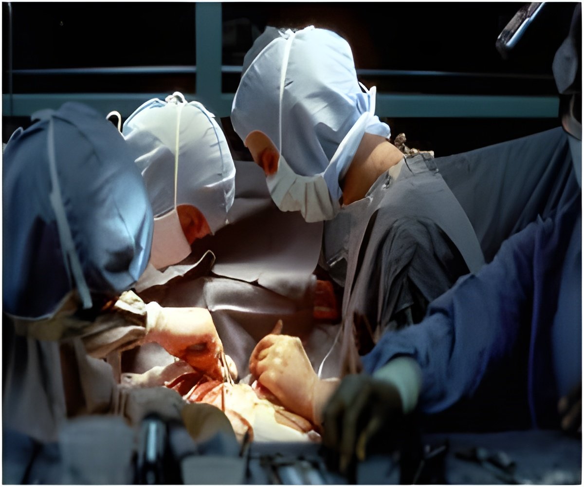
Using transesophageal echocardiography (TEE) during heart surgery may improve outcomes for children with congenital heart disease.

‘Using cardiac imaging during heart surgery can detect residual holes in the heart that may occur when surgeons repair a child's heart defect, and offers an opportunity to close those holes during the same operation.’


Using cardiac imaging during heart surgery can detect serious
residual holes in the heart that may occur when surgeons repair a
child's heart defect, and offers surgeons the opportunity to close those
holes during the same operation. Pediatric cardiology experts say using
this tool, called transesophageal echocardiography (TEE), during
surgery may improve outcomes for children with congenital heart disease.
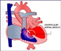
"We focused on intramural ventricular septal defects, which are holes between two chambers of the heart," said Meryl S. Cohen, senior author and pediatric cardiologist at Children's Hospital of Philadelphia (CHOP). She and co-authors previously published a paper in Circulation that recognized these defects as being distinct from other types of residual holes.
"These defects, which can occur after initial surgery for another defect, can increase the risk of complications and mortality in children with heart disease, so using imaging tools to quickly identify these defects can improve our care of these children," she added.
The study's first author, Jyoti K. Patel was a former cardiac fellow in the Cardiac Center at CHOP, and conducted the research during her fellowship. The study team published the research in the September 2016 issue of the Journal of Thoracic and Cardiovascular Surgery.
The scientists reported on the use of intraoperative TEE to identify intramural ventricular septal defects (VSDs). They performed a retrospective study of 337 children, mostly infants, who underwent surgery at CHOP for conotruncal defects from 2006 to 2013.
Advertisement
Of the 337 surgical patients, 34 had intramural VSDs. Of those 34, both TTE and TEE identified 19 VSDs, while 15 were identified by TTE only. That data showed that TEE had modest sensitivity (56 percent), but high specificity (100 percent) in identifying intramural VSDs. The authors note that "the modest sensitivity suggests that many intramural defects are not detected in the operating room." However, they add, intraoperative TEE was able to identify most of the intramural defects requiring reintervention (e.g., further surgery).
Advertisement
Source-Eurekalert


