Survival and muscle function in rats used to model ALS, improved upon transplantation of human stem cells in an experiment conducted at the University of Wisconsin-Madison.
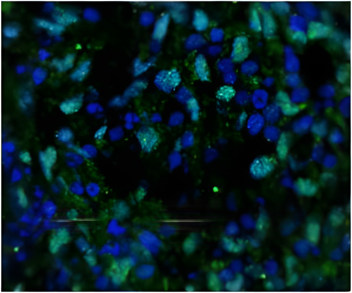
In work recently completed at the UW School of Veterinary Medicine, Masatoshi Suzuki, an assistant professor of comparative biosciences, and his colleagues used adult stem cells from human bone marrow and genetically engineered the cells to produce compounds called growth factors that can support damaged nerve cells.
The researchers then implanted the cells directly into the muscles of rats that were genetically modified to have symptoms and nerve damage resembling ALS.
In people, the motor neurons that trigger contraction of leg muscles are up to three feet long. These nerve cells are often the first to suffer damage in ALS, but it's unclear where the deterioration begins. Many scientists have focused on the closer end of the neuron, at the spinal cord, but Suzuki observes that the distant end, where the nerve touches and activates the muscle, is often damaged early in the disease.
The connection between the neuron and the muscle, called the neuro-muscular junction, is where Suzuki focuses his attention. "This is one of our primary differences," Suzuki says. "We know that the neuro-muscular junction is a site of early deterioration, and we suspected that it might be the villain in causing the nerve cell to die. It might not be an innocent victim of damage that starts elsewhere."
Previously, Suzuki found that injecting glial cell line-derived neurotropic factor (GDNF) at the junction helped the neurons survive. The new study, published in the journal Molecular Therapy on May 28, expands the research to show a similar effect from a second compound, called vascular endothelial growth factor.
Advertisement
But the real advance, Suzuki says, was finding an even better result from using stem cells that create both of these two growth factors. "In terms of disease-free time, overall survival, and sustaining muscle function, we found that delivering the combination was more powerful than either growth factor alone. The results would provide a new hope for people with this terrible disease."
Advertisement
The injected stem cells survived for at least nine weeks, but did not become neurons. Instead, their contribution was to secrete one or both growth factors.
Originally, much of the enthusiasm for stem cells focused on the hope of replacing damaged cells, but Suzuki's approach is different. "These motor nerve cells have extremely long connections, and replacing these cells is still challenging. But we aim to keep the neurons alive and healthy using the same growth factors that the body creates, and that's what we have shown here."
For the test, Suzuki used ALS model rats with a mutation that is found in a small percentage of ALS patients who have a genetic form of the disease. "This model has been accepted as the best test bed for ALS experiments," says Suzuki.
By using adult mesenchymal stem cells, the technique avoided the danger of tumor that can arise with the transplant of embryonic stem cells and related "do-anything" cells. Importantly, mesenchymal stem cells have been already used in clinical trials for various human diseases.
In the future, Suzuki hopes to apply his approach by using clinical grade stem cells. "Because this is a fatal and untreatable disease, we hope this could enter a clinical trial relatively soon."
Source-Eurekalert


