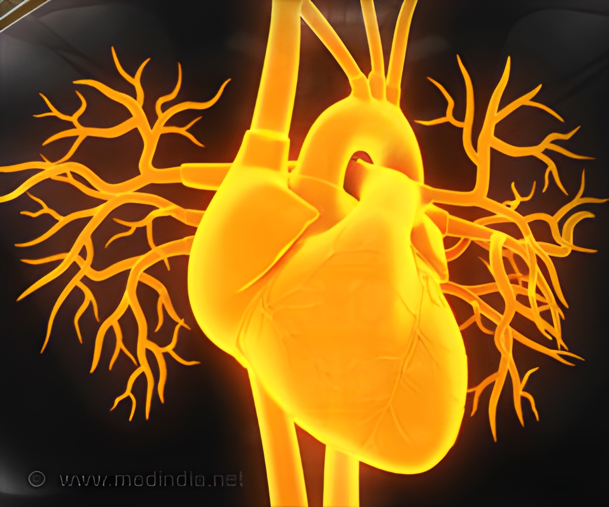
High eosinophil levels in EHD cause heart damage through specific cell death pathways.

Eosinophils and Heart Damage in EHD
The study published in AHA Journals, Circulation, tested in vitro to find that eosinophils undergo regulated necrosis and damage cardiomyocytes. The research was conducted in a mouse model of EHD (1✔ ✔Trusted SourceEosinophil Regulated Necrosis in Murine Model of Eosinophilic Heart Disease
Go to source).
The mice model was administered with high levels of eosinophil and cardiac myosin to stimulate the mice’s immune system. Specific biomarkers and tissues were analyzed to see how the disease develops.
EHD mice showed high levels of eosinophils in the heart, causing cardiomyocyte necrosis and later fibrosis. However, heart inflammation was decreased in eosinophil-deficient mice. Due to necrosis, the release of cell-free DNA and troponin was detected in the plasma.
Eosinophil Cell Death Pathways in Heart Disease
When an immunohistochemistry test was done for citH3, a marker of regulated necrosis, showed eosinophils in the tissue that were undergoing regulated necrosis. To study regulated necrosis of eosinophils in vitro, eosinophils were stimulated with known to induce different types of cell death and stimuli that mimic activation by conditions in the inflammatory microenvironment.While all the stimuli induced time-dependent death, two different patterns were observed. Some stimuli like CD95 ligation caused eosinophils to transition through the annexinV+/7AAD- stage (early apoptosis) and other stimuli (e.g. PMA and ionomycin) had an increased ratio of 7AAD+ cells. This indicates that they directly enter the necrotic pathway.
Advertisement
This study shows that mouse eosinophils undergo a specific type of cell death pathway, and this may have different effects on disease outcomes.
Advertisement
- Eosinophil Regulated Necrosis in Murine Model of Eosinophilic Heart Disease - (https://www.ahajournals.org/doi/10.1161/circ.150.suppl_1.4141864)
Source-Medindia


