A routinely used invasive heart test to measure heart function is being severely overused, particularly among patients who have recently undergone similar, more effective tests, states a new study.

The procedure, called left ventriculography or left ventriculogram, was developed 50 years ago to assess how well the heart functions by using a measurement method called "ejection fraction" — the percentage of blood that gets squeezed out with each heartbeat. The investigators found that it is routinely performed as an add-on procedure during a coronary angiogram, a separate heart-imaging test, at an extra cost of $300.
Over the years, several less-invasive and often superior methods of measuring ejection fraction have emerged, such as echocardiograms and nuclear cardiac imaging, making the use of left ventriculography questionable at times, the study states.
The study appears online this month in the American Heart Journal.
Several years ago when Witteles was a cardiac fellow, he and his colleagues noticed a great deal of variation in whether cardiologists would order the procedure, often in similar patient cases, he said. This seemingly arbitrary use of left ventriculography led to the idea for this study.
Researchers first set out to determine exactly how often the procedure was conducted. They examined a national database of about 96,000 patients enrolled in Aetna health benefits plans in 2007 who underwent a coronary angiogram during that year. The data showed left ventriculography was performed 81.8 percent of the time whenever an angiogram was done — a surprisingly high rate, Witteles said.
Advertisement
"If a patient recently had an echocardiogram or a nuclear study, it didn't make them less likely to have the left ventriculography procedure — it made them more likely," Witteles said. "That is impossible to explain from a medical justification standpoint.
Advertisement
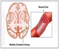
Even more concerning than the added costs are the medical risks from performing an unnecessary procedure. For left ventriculography, this can include side effects from injecting contrast dye (which can be particularly harmful for patients with kidney dysfunction or diabetes), increased radiation exposure and an increased risk of abnormal heart rhythms and stroke.
During a coronary angiogram, a catheter is threaded through the blood vessels to the heart, contrast dye is inserted and X-rays are taken. The add-on left ventriculography procedure involves moving the catheter across the aortic valve of the heart and inserting another dose of contrast dye. This allows visualization of the left ventricle and its contractions.
"The biggest downside is that the catheter goes across the valve into the heart," Witteles said. "There's always a risk of dislodging a blood clot, causing a stroke. The procedure only takes five minutes, but it increases the risk of arrhythmias. And then there is the added cost. But the real big-picture issue is how often an unnecessary, invasive test is being routinely ordered."
Source-Eurekalert


