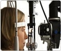
Eye ultrasounds provide a swift and secure means to detect brain shunt failure in children, facilitating crucial diagnosis and treatment in emergency scenarios.

‘Eye ultrasounds offer a rapid and safe method to identify #brainshunt failure in children, aiding timely diagnosis and treatment in emergency settings. #childhealth #eyeultrasound’


Identifying Shunt Failure in Emergency Settings
When a patient visits the emergency department for potential shunt failure, their symptoms are often nonspecific, including headache, vomiting, and fatigue, according to researchers. Shunt failure is life threatening, and children with shunts typically undergo multiple computed tomography and magnetic resonance imaging scans per year, exposing them to excessive radiation and sedation. A backup of fluid causes the optic nerve sheath to swell, which researchers can measure with eye ultrasound.The study found that comparing the diameter of the optic nerve when a patient is symptomatic to the diameter when they are well can help determine if a shunt is blocked.
“The research team is interested in finding ways to lessen radiation exposure and expedite diagnosing shunt failure in the emergency department,” said Adrienne L. Davis, MD, MSc, FRCPC, pediatric emergency medicine research director at The Hospital for Sick Children (SickKids) and presenting author. “The study uses patients as their own controls by measuring the optic nerve when well and sick—a strategy that individualizes this test for every patient and recognizes that every patient with a shunt has a unique degree of shunt dependence and ability to tolerate high brain pressures.”
The researchers studied 76 pairs of eye ultrasounds of nearly 60 children presenting to the Toronto hospital’s emergency department with potential shunt failure. Researchers note that while findings are promising, results require further confirmation in a larger population of children with shunts across North America.
Reference:
- CHange in Optic nerve sheath diameter (ONSD) and Optic disc elevation (ODE) in predicting Shunt failure in the Emergency department - (https://www.pas-meeting.org/)


