Molecular and protein markers that appear soon after liver transplant in hepatitis C patients could be a sign of failure of the transplant and predict rapid onset of liver damage.
The findings were the cover story of the liver disease journal Hepatology. The molecular and protein markers were found in separate studies, one led by Dr. Angela "Angie" Rasmussen, and the other by Dr. Deborah Diamond, both working in the lab of Dr. Michael Katze, professor of microbiology at the University of Washington. The Katze lab employs systems approaches to studying many viral pathogens, including hepatitis C, HIV, influenza, SARS and Ebola.
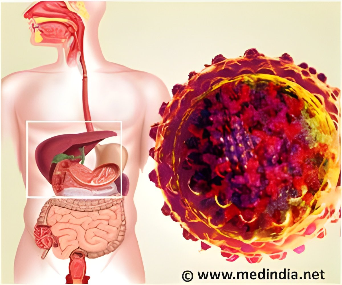
Liver failure from chronic hepatitis C infection is the leading reasons for liver organ transplantation surgery worldwide, Rasmussen noted. In almost all cases, the newly transplanted organ quickly becomes infected, because the hepatitis C virus persists in the patient's bloodstream. Virus damage to the transplanted organ is sometimes slow, taking years or decades to progress.
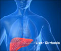
However, in about a third of the cases, she said, leathery, fibrous tissue builds up in the transplanted liver within one to two years. This condition, called cirrhosis, can keep the liver from clearing toxins from the body and can end in liver transplant failure.
Physicians have had no means of sorting out which of their patients might be prone to this complication. They have been awaiting prognostic markers for disease progression and therapy response to allow them to tailor treatment to the individual patient.
Rasmussen said that, at present, liver patients regularly have invasive core-needle biopsies to check their transplanted liver. Strong antiviral drugs are available to try to offset hepatitis C liver damage, she explained, but these are not always effective and usually have harsh side effects. Surgeons have been hoping for a test that could indicate if a patient was likely to do well with less aggressive treatment. This knowledge might spare the patient from unnecessary repeat biopsies and the most potent antiviral regimens, which impose severe, flu-like discomfort, chronic fatigue, depression and anemia.
Advertisement
The two research teams led by Rasmussen and Diamond took different, highly sophisticated approaches to find prognostic markers. The Diamond group performed global protein analyses of liver biopsies taken at 6 months and a year after transplantation. State-of-the art mass spectrometry methods and computational modeling directed them to 250 proteins, out of 4,324 originally uncovered, whose regulation was markedly different in patients with rapidly progressing fibrosis. These patients showed an enrichment of regulatory proteins associated with various immune, liver-protective, and fibrosis-generating processes. The researchers also observed an increase in proinflammatory activity and impairment in antioxidant defenses.
Advertisement
Rasmussen's team faced the challenge that no single clinical variable, or combination of clinical variables, could accurately warn that a liver transplant patient was heading for trouble. They decided to look at all of the RNA molecules produced in cells from 111 liver biopsy specimens collected over time from 57 hepatitis C infected patients who had undergone liver transplant surgery. Such a set of molecules is called a transcriptome, a collection of works "written" from the DNA code in response to a viral infection. Her team applied leading-edge mathematical modeling systems to cull patterns of gene expression.
"We were able to identify a molecular signature of gene expression for those patients at risk of developing severe fibrosis in their transplanted livers," Rasmussen said.Significantly, her team also discovered that these alterations in gene expression occur before there is evidence in the liver tissue of disease progression.
"That suggested to us that events that occur during the early stages when the transplanted liver is becoming infected with the patient's hepatitis C virus influence the course of the disease," Rasmussen said. Her team also observed a precursor state – a set of conditions that foretold problems ahead – that was common for different, severe clinical outcomes. Her team went on to describe the probable cellular network that could be the basis for the initial transition to severe liver disease.
They hope the findings open new avenues in research to delay disease progression and extend the survival of transplanted livers. Both the Rasmussen and the Diamond teams employed systems biology methods to understand the root of post-transplant liver disease recurrence. This approach looks at the complex interactions between pathogens, in this case the hepatitis C virus, and the organism they infect. These interactions are largely studied in their entirety at the scale of the genetic, protein and cellular alterations that occur.
There study results are a first for systems biology research: they revealed molecular markers associated with disease outcome, not simply markers of hepatitis C infection. The researchers' groundbreaking scientific approach to the effects of viral infection, as well as their findings, were praised in an accompanying editorial in Hepatology.
Source-Eurekalert

