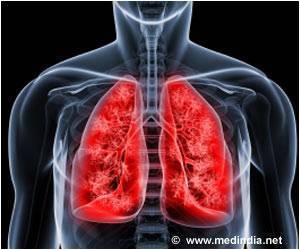
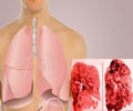
Pulmonary fibrosis is caused by scarring that seems to feed on itself, and is incurable. Tougher less elastic tissue replaces the stretchable lung, making breathing difficult.
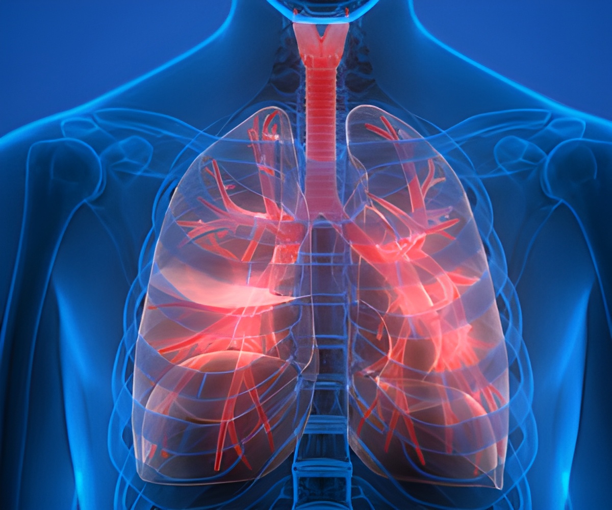
"In the advanced stages of the disease, there’’s not a lot we can do for patients," says Dr. Summer. Some existing therapies alleviate symptoms, but none reverse or stop disease progression. Many patients live only three to five years after diagnosis, suggests the American Lung Association and the only effective treatment is lung transplant. The team led by Dr. Summer and first author Freddy Romero, Ph.D., looked at a mouse model of lung fibrosis initiated by a chemical known to cause the disease. Researchers noticed that lipids (AKA fat), accumulated within the airspaces of the lung where oxygen is absorbed. Although lipids are normally secreted there to help keep the cells lining the lungs lubricated and properly inflated, these were excessive levels of fat.
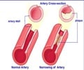
The researchers showed that in response to stress, the cells producing the lubricant dump their lipid stores into the lungs and fail to mop up the excess. The excess lipids react with oxygen to create a form of fat that acts as an inflammatory signal; in some ways this response is similar to the events that initiate atherosclerosis, or plaque formation in blood vessels. In the lungs, Dr. Summer’’s laboratory showed that immune cells called macrophages, which normally survey the lung for debris, infection, or dying cells begin gobbling up the excess fat in the lungs. Loaded with this oxidized fat, the macrophages turned on a program that acts to help heal the wounded tissue, but as a consequence to this adaptive response leads to the development of fibrotic lung disease.
"Both the initial damage to the cells lining the airway of the lung and the inflammation are important," says Dr. Romero, "but the thing that drives the damage is the unregulated excess lipids in the distal airspaces." When the researchers put oxidized lipids into the lungs of mice that had not been exposed to any lung-damaging chemicals, the mice also developed fibrosis, showing that the oxidized fat alone was enough to cause the disease.
"These results show, for the first time, that a break-down of normal lipid handling may be behind this lung disease," says Dr. Summer "If we prove that the same process holds true in humans, it suggests that we could prevent or mitigate the disease by simply clearing out the excess oxidized lipids from lungs."
To this end, the researchers tested whether treating mice with an agent called GM-CSF that reduces lipid secretion and facilitates lipid removal in the lungs, could minimize lung fibrosis. Indeed, this agent reduced the scarring in the lungs by over 50 percent based on the levels of lung collagen, a marker of newly forming scar tissue. In addition, the researchers examined human cells in the lab and saw that oxidized fat also promoted a fibrotic response.
Advertisement
Research was supported by funding from the National Institutes of Health (NIH) R01HL105490 and by the Intramural Research Program of the National Institutes of Health, National Institute of Environmental Health Sciences grant Z01 ES102005.
Advertisement
About Jefferson - Health is all we do.
Thomas Jefferson University, Thomas Jefferson University Hospitals and Jefferson University Physicians are partners in providing the highest-quality, compassionate clinical care for patients, educating the health professionals of tomorrow, and discovering new treatments and therapies that will define the future of healthcare.
Thomas Jefferson University enrolls more than 3,600 future physicians, scientists and healthcare professionals in the Sidney Kimmel Medical College (SKMC); Jefferson Schools of Health Professions, Nursing, Pharmacy, Population Health; and the Graduate School of Biomedical Sciences, and is home of the National Cancer Institute (NCI)-designated Sidney Kimmel Cancer Center. Jefferson University Physicians is a multi-specialty physician practice consisting of over 650 SKMC full-time faculty.
Thomas Jefferson University Hospitals is the largest freestanding academic medical center in Philadelphia. Services are provided at five locations - Thomas Jefferson University Hospital and Jefferson Hospital for Neuroscience in Center City Philadelphia; Methodist Hospital in South Philadelphia; Jefferson at the Navy Yard; and Jefferson at Voorhees in South Jersey.
Source-Newswise

