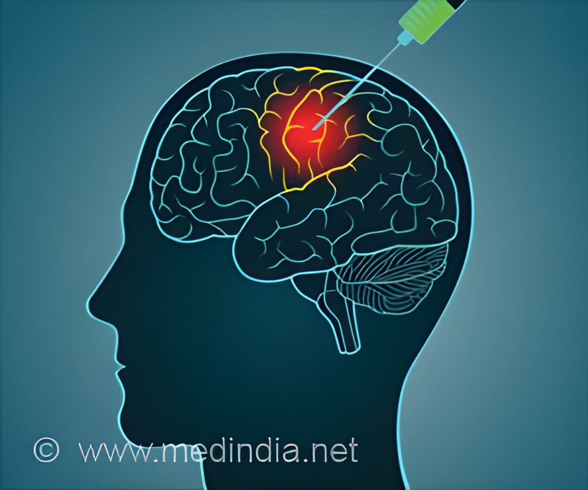
Scientists unveil the first-ever neuron map of the adult brain, offering groundbreaking insights into brain function and paving the way for future neurological research.

Whole-brain annotation and multi-connectome cell typing of Drosophil
Go to source). This landmark achievement has been conducted by a large international collaboration of scientists, called the FlyWire Consortium, including researchers from the MRC Laboratory of Molecular Biology (in Cambridge, UK), Princeton University, the University of Vermont and the University of Cambridge. It is published in a pair of papers in Nature.
‘Scientists have mapped the entire #brain of a #fruitfly, neuron by #neuron! This breakthrough could unlock secrets of larger brains, including our own. #nervousdisorder’


A Giant Leap in Brain Mapping
The diagram of all 139,255 neurons in the adult fly brain is the first of an entire brain for an animal that can walk and see. Previous efforts have completed the whole brain diagrams for much smaller brains, for example, for that of a fruit fly larva, which has 3,016 neurons, and a nematode worm, which has 302 neurons.Dr. Gregory Jefferis, from the MRC Laboratory of Molecular Biology and from the University of Cambridge, who was one of the co-leaders of the research, said: “If we want to understand how the brain works, we need a mechanistic understanding of how all the neurons fit together and let you think. For most brains we have no idea how these networks function."
“Flies can do all kinds of complicated things like walk, fly, navigate, and the males sing to the females. Brain wiring diagrams are a first step towards understanding everything we’re interested in – how we control our movement, answer the telephone, or recognise a friend.”
Dr Mala Murthy, from Princeton University, who was one of the co-leaders of the research, said: “We have made the entire database open and freely available to all researchers. We hope this will be transformative for neuroscientists trying to better understand how a healthy brain works. In the future we hope that it will be possible to compare what happens when things go wrong in our brains, for example in mental health conditions.”
The scientists found that there were substantial similarities between the wiring in this map and previous smaller-scale efforts which have mapped out parts of the fly brain. This led the researchers to conclude that there are many similarities in wiring between individual brains – that each brain isn’t a unique structure like a snowflake.
Advertisement
A whole fly brain is less than 1 millimetre wide. The researchers started with one female brain cut into seven thousand slices, each only 40 nanometres thick, that were previously scanned using high resolution electron microscopy in the laboratory of project co-leader Davi Bock, then at Janelia Research Campus in the US.
Advertisement
However, the AI still makes many errors in datasets of this size. The FlyWire Consortium – made up of teams in more than 76 laboratories and 287 researchers around the world, as well as volunteers from the general public – spent an estimated 33 person-years painstakingly proofreading all the data.
Dr Sebastian Seung, from Princeton University, who was one of the co-leaders of the research, said: “Mapping the whole brain has been made possible by advances in AI computing - it would have not been possible to reconstruct the entire wiring diagram manually. This is a display of how AI can move neuroscience forward. The fly brain is a milestone on our way to reconstructing a wiring diagram of a whole mouse brain.”
The researchers also annotated many details on the wiring diagram, such as classifying more than 8,000 cell types across the brain. This also allows researchers to select particular systems within the brain for further study, such as the neurons involved in sight or movement.
Dr. Philipp Schlegel, the first author of one of the studies, from the MRC Laboratory of Molecular Biology, said: “This dataset is a bit like Google Maps but for brains: the raw wiring diagram between neurons is like knowing which structures on satellite images of the earth correspond to streets and buildings. Annotating neurons is like adding the names for streets and towns, business opening times, phone numbers, reviews, et cetera to the map – you need both for it to be really useful.”
This is also the first whole brain wiring map – often called a connectome – to predict the function of all the connections between neurons.
Neurons use electrical signals to send messages. Each neuron can have hundreds of branches that connect it to other neurons. The points where these branches meet and transmit signals between neurons are called synapses. There are two main ways that neurons communicate across synapses: excitatory (which promotes the continuation of the electrical signal in the receiving neuron), or inhibitory (which reduces the likelihood that the next neuron will transmit signals).
Researchers from the team also used AI image scanning technology to predict whether each synapse was inhibitory or excitatory.
Dr. Gregory Jefferis added: “To begin to simulate the brain digitally, we need to know not only the structure of the brain, but also how the neurons function to turn each other on and off.”
“Using our data, which has been shared online as we worked, other scientists have already started trying to simulate how the fly brain responds to the outside world. This is an important start, but we will need to collect many different kinds of data to produce reliable simulations of how a brain functions.”
Associate Professor Davi Bock, who was one of the co-leaders of the research, from the University of Vermont, said: “The hyper-detail of electron microscopy data creates its own challenges, especially at scale. This team wrote sophisticated software algorithms to identify patterns of cell structure and connectivity within all that detail."
“We now can make precise synaptic level maps and use these to better understand cell types and circuit structure at whole-brain scale. This will inevitably lead to a deeper understanding of how nervous systems process, store and recall information. I think this approach points the way forward for the analysis of future whole-brain connectomes, in the fly as well as in other species."
This research was conducted using a female fly brain. Since there are differences in neuronal structure between male and female fly brains, the researchers plan to also characterise a male brain in the future.
The principal funders were the National Institutes of Health BRAIN Initiative, Wellcome, Medical Research Council, Princeton University, and National Science Foundation.
Reference:
- Whole-brain annotation and multi-connectome cell typing of Drosophila - (https://www.nature.com/articles/s41586-024-07558-y)


