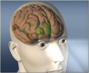
US researchers are reporting success in getting through blood-brain barrier for tumor imaging and treatment. The barrier protects the brain from the bloodstream, thus inhibiting drug delivery.

In experiments with mice, the researchers demonstrated that the targeted particles guide payloads to image tumors, treat tumors, or can potentially do both to monitor treatment as it occurs.
“We’ve identified an iron-mimic peptide that can hitch a ride on a protein complex that transports iron across the blood-brain barrier,” said co-senior author Wadih Arap, M.D., Ph.D., professor in the David H. Koch Center at MD Anderson. “Employing the iron transport system selectively opens the blood-brain barrier for tumor imaging and treatment while keeping it otherwise intact to play its protective role.”
The barrier thwarts drug delivery because its tight layering of blood vessel cells and certain types of brain cells forms a nearly impenetrable wall against most blood-borne compounds, which can harm the brain. The iron-transporting transferrin protein and receptor complex is a potential path to treatment, the authors noted, because its receptor gene is the most overexpressed in human glioblastomas.
Glioblastomas are the most common form of primary brain tumor among adults and are one of the most lethal cancers — patients have a median survival rate of one year. They resist radiation and chemotherapy and often invade areas of the brain where full surgical removal is impossible.
Arap and Renata Pasqualini, Ph.D., professor in the Koch Center and co-senior author on the paper, developed the screening, targeting and delivery processes used in the project. The combination consists of a viral particle called phage packaged with a bit of protein, or peptide, which acts as a ligand, binding to the target.
Advertisement
The team is working toward phase I human clinical trials, but required preclinical steps will take at least two years. “Our priority is the rapid translation of our approach into clinical applications, an effort that will greatly benefit from the collaborative, multidisciplinary nature of this project,” said co-first author Fernanda Staquicini, Ph.D., a postdoctoral fellow in the Arap-Pasqualini lab.
Advertisement
The iron-binding protein tranferrin has two conformations, one when it’s iron-free (open) and another when it contains iron (closed). Experiments showed that CRTIGPSVC connects with the open form, converting the protein to a state similar to the closed form, as if it were carrying iron. The closed form binds to transferrin receptors, which exist in abundance in glioblastoma.
The CRTIGPSVC peptide phage was given to mice with human-derived malignant glioma. An hour later, the tumors harbored high levels of the peptide phage, which appeared at only background levels in normal tissue.
Immunostaining showed the peptide phage gathered in both the tumor’s blood vessels and tumor tissue, indicating that it passed from the blood vessels to tumor cells.
The CRTIGPSVC peptide was then connected to a viral delivery system loaded with a gene from the Herpes’ simplex virus known as HSVtk. It serves as a reporter gene for molecular imaging with PET scans and causes cells to kill themselves when given with the drug ganciclovir.
Mice treated with the CRTIGPSVC-targeted versions loaded with the HSVtk gene had tumors that were typically half the size of those in mice treated with the untargeted delivery system when both groups were treated with ganciclovir.
Blood vessel and glioma cells forced to commit suicide (apoptosis) were found throughout the tumors of mice treated with the peptide-targeted delivery system. Cell suicide did not occur above background levels in normal brain tissue.
PET/CT scans showed widespread targeting of tumor tissue with little or no signal in normal tissue.
The team analyzed expression of the transferrin receptor in 165 samples of human tumors. A strong or moderate presence of the receptor was found on 85 percent of glioblastoma samples, indicating that the receptor might be a suitable target for human use.
Peptide-guided delivery of drugs or imaging agents could apply to other diseases of the central nervous system, said co-author Richard Sidman, M.D., emeritus professor of neuropathology at Harvard Medical School and the Department of Neurology at Beth Deaconess Medical Center.
“One important set of diseases to test will be lysosomal storage diseases, mostly lethal in childhood or adolescence, in each of which a different crucial brain enzyme is markedly reduced or absent because of a defect in the corresponding gene,” Sidman said. “These diseases can be treated by provision to the brain of 25 percent or less of the missing enzyme. Therefore, the prospect merits testing as to whether delivery of even modest amounts of the normal version of the pertinent gene across the blood-brain barrier would be therapeutically effective.”
Prospects for radiologically diagnosing and treating strokes, traumatic injury and neurodegenerative diseases such as Alzheimer’s, Parkinson’s, and Huntington’s diseases, also are worth pursuing, Sidman noted.
An accompanying commentary in JCI notes the flexibility and utility of the researchers’ plan of attack. “We can look forward to future applications of this approach for the discovery of new druggable targets and the identification of cell surface molecules that may be important in many types of cancer as well as other diseases,” concluded co-authors David Nathanson, M.D., and Paul Mischel, M.D., of the David Geffen School of Medicine at UCLA.
Source-Medindia


